

3D Model of Mandibular First Molar Anatomy 3D print model
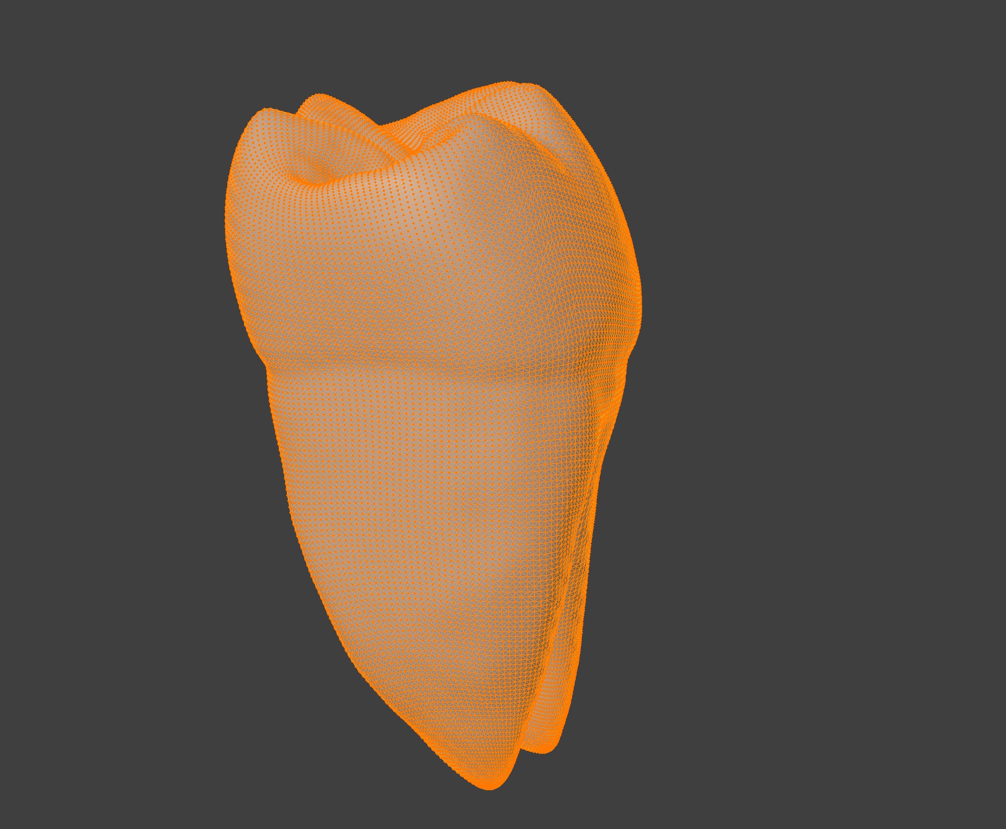
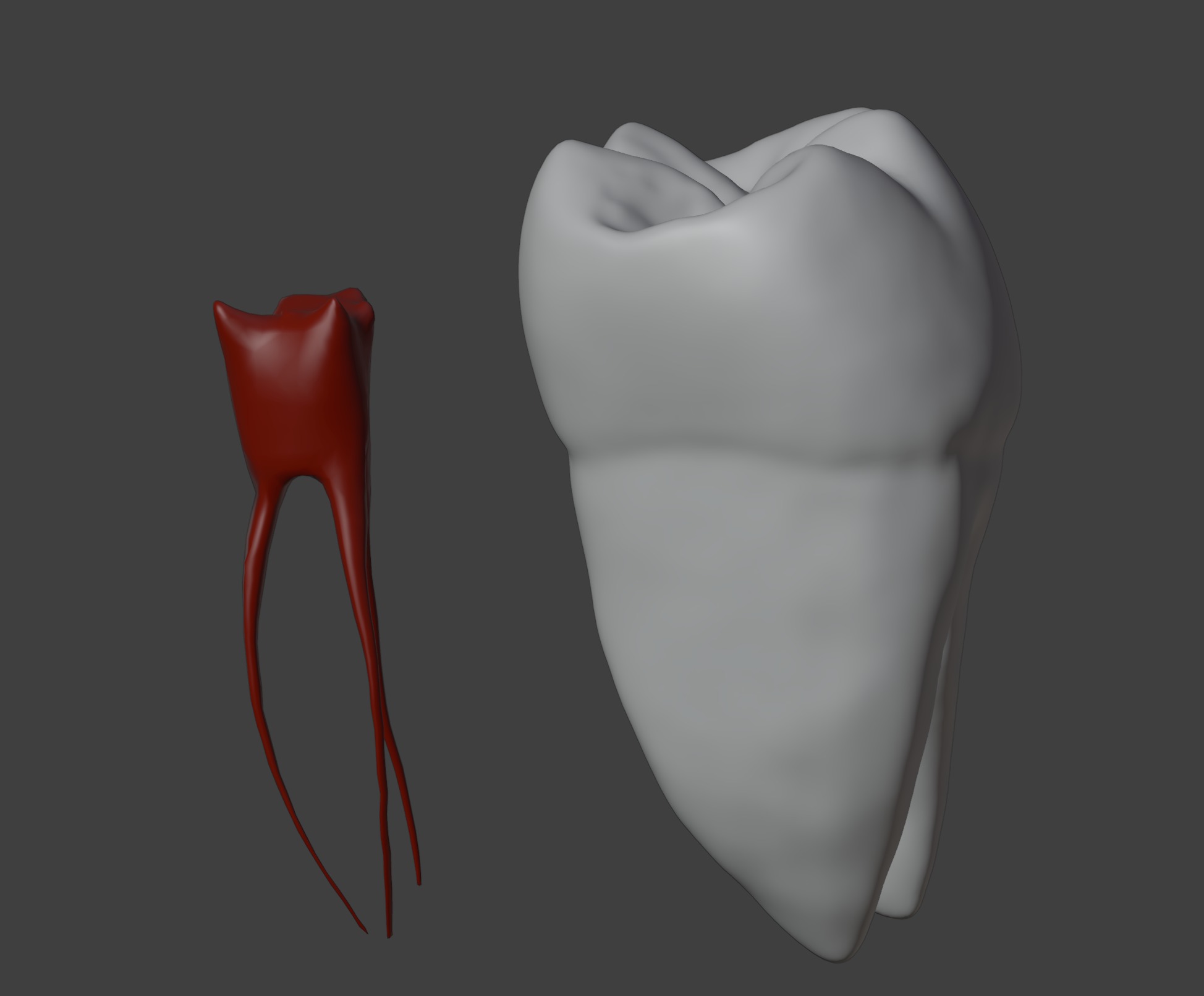
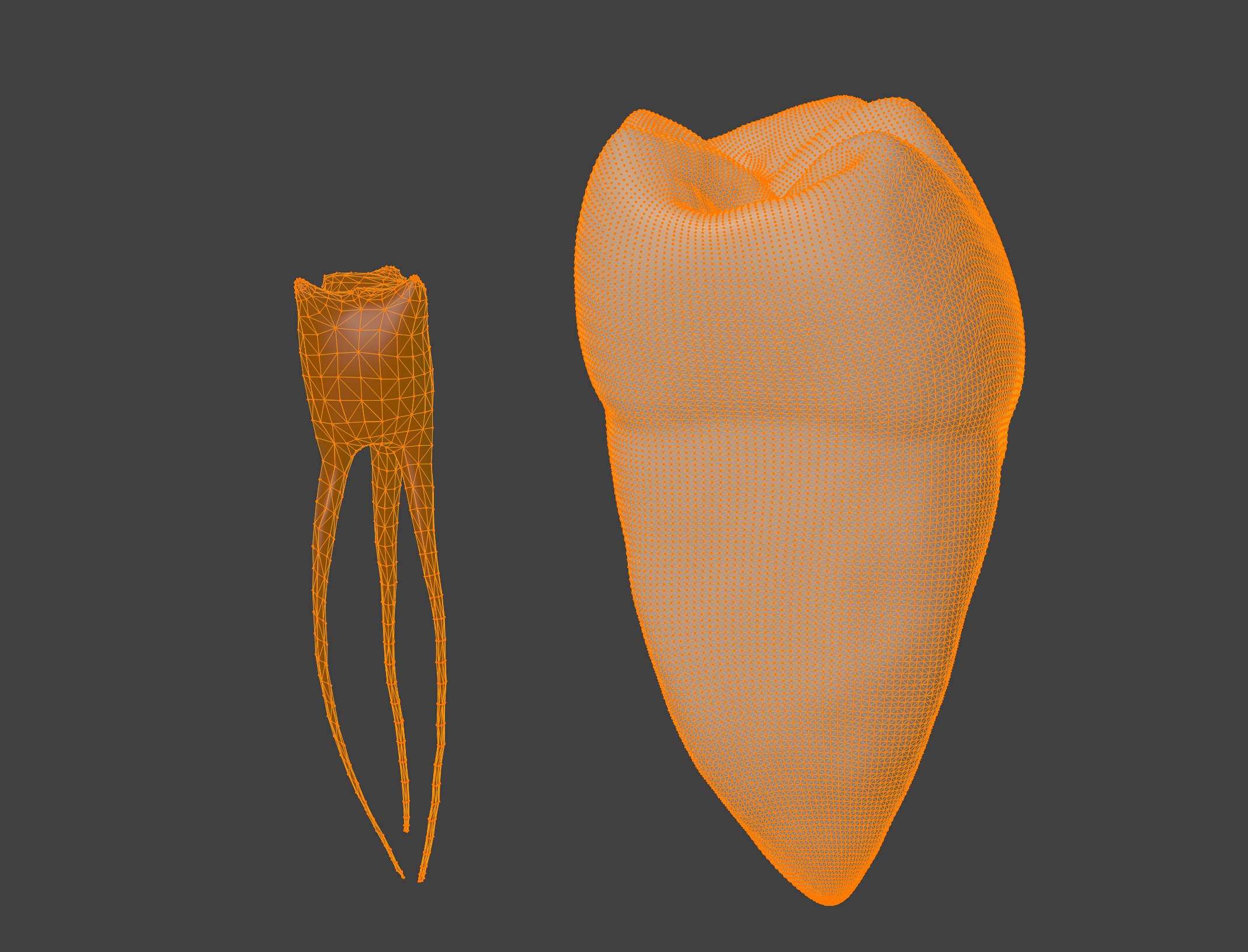
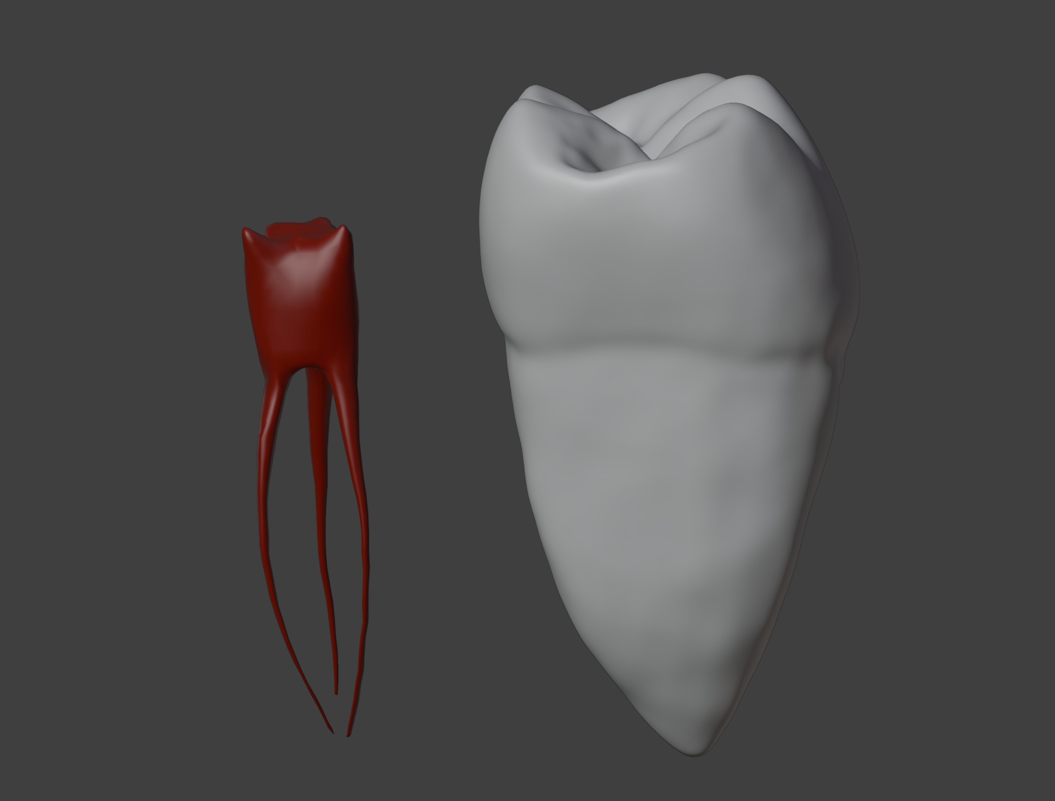
This 3D model of mandibular first molar anatomy provides a highly detailed and anatomically accurate representation of the structure and function of a lower first molar tooth. It highlights key features, including the enamel, dentin, pulp chamber, root canals, and cusps, offering a clear visualization of both internal and external anatomy. The model also showcases the two distinct roots and multiple cusps designed for efficient grinding and chewing of food, demonstrating the molar's critical role in mastication and digestion.
Designed for educational and clinical purposes, this model is an invaluable tool for studying dental anatomy, understanding root morphology, and visualizing conditions such as cavities, fractures, root canal infections, and periodontal diseases. Ideal for dentists, endodontists, educators, and students, it supports teaching, training, and patient education by providing a hands-on approach to exploring the structure, function, and care of mandibular molar teeth.

3D Model formats
Format limitations
- glTF (.gltf, .glb)1.25 MB
- Collada (.dae)7.78 MB
- OBJ (.obj, .mtl) (2 files)4.95 MB
- Ply (.ply)1.29 MB
- Stereolithography (.stl)2.72 MB
- Blender (.blend)5.2 MB
3D Model details
- Ready for 3D Printing
- Publish date2025-01-06
- Model ID#5759431
Similar Models

Users who bought this item also bought...
