


3D Model of Lower Single-Root Pre-Molar Tooth 3D print model
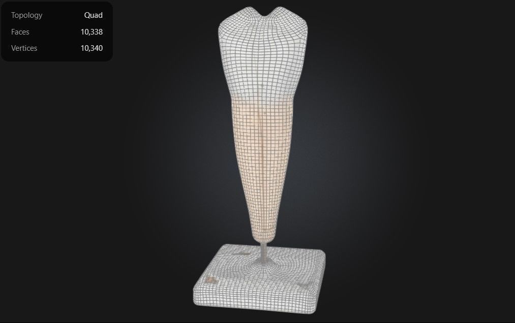
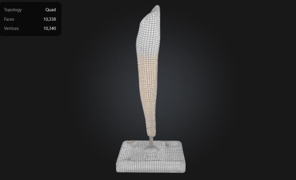
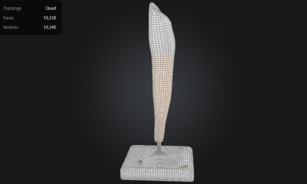
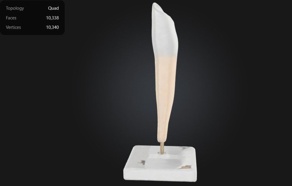
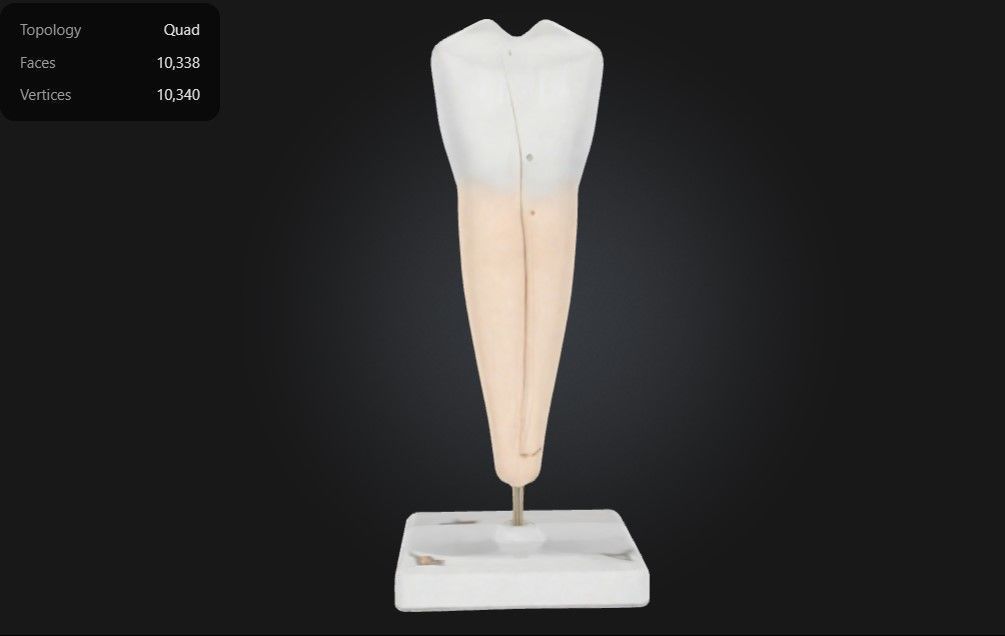
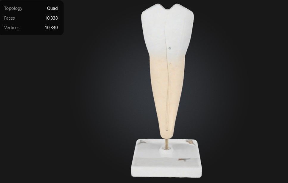
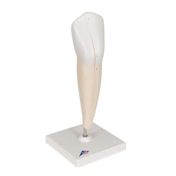
This 3D model of a lower single-root premolar tooth provides a highly detailed and anatomically accurate representation of the structure and function of a mandibular premolar. It highlights essential components, including the enamel, dentin, pulp chamber, and root canal, offering a clear view of the tooth’s internal and external anatomy.
The model also showcases the crown’s cusps and ridges, designed for both tearing and grinding food, and emphasizes the single-root structure that anchors the tooth securely in the jawbone.
Designed for educational and clinical purposes, this model is an invaluable tool for studying dental anatomy, understanding root morphology, and visualizing conditions such as cavities, fractures, and periodontal diseases.
Ideal for dentists, endodontists, educators, and students, it supports teaching, training, and patient education by providing a hands-on approach to learning about the structure, function, and care of premolar teeth.

3D Model formats
Format limitations
- Stereolithography (.stl)1010 KB
- OBJ (.obj, .mtl)2.57 MB
- PNG (.png) (2 files)7.56 MB
- Autodesk FBX (.fbx)4.99 MB
- glTF (.gltf, .glb)1.05 MB
- Blender (.blend)3.58 MB
- USDZ (.usdz)935 KB
3D Model details
- Ready for 3D Printing
- Publish date2025-01-05
- Model ID#5757786
Similar Models

Users who bought this item also bought...
