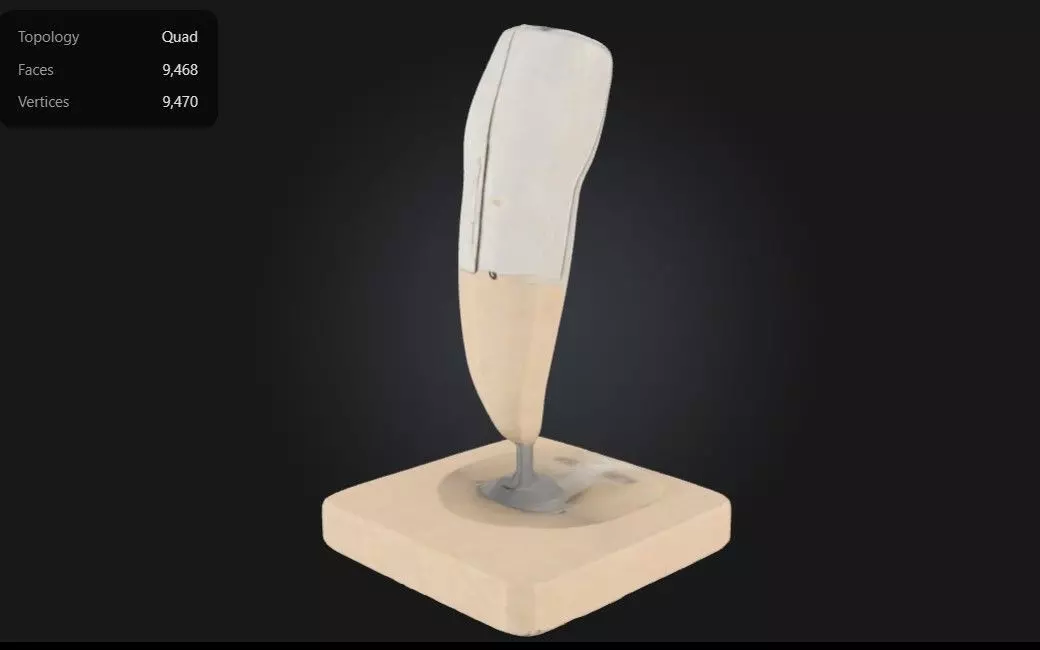
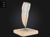
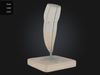
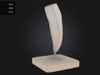
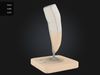
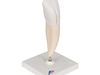
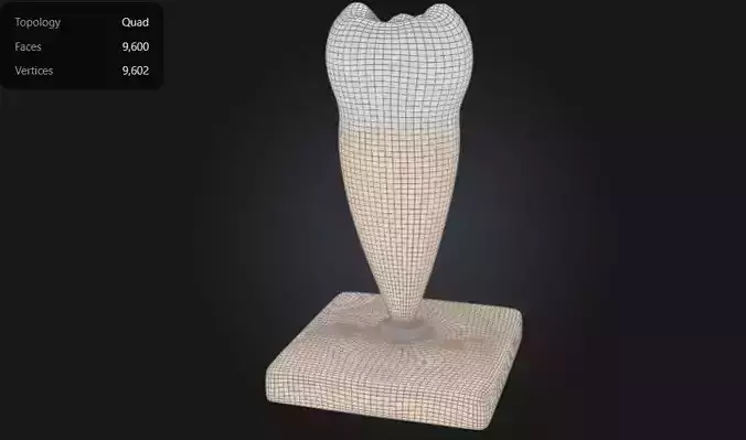
This 3D model of a lower incisor tooth provides a highly detailed and anatomically accurate representation of the structure and function of a mandibular incisor. It highlights key features, including the enamel, dentin, pulp chamber, and root canal, offering a clear visualization of the tooth’s internal and external anatomy.
The model also showcases the thin, chisel-shaped crown designed for cutting and slicing food, along with its single, narrow root that provides stability within the jawbone.
Designed for educational and clinical purposes, this model is an excellent tool for studying dental anatomy, understanding root morphology, and visualizing conditions such as cavities, fractures, and periodontal diseases.
Ideal for dentists, orthodontists, educators, and students, it supports teaching, training, and patient education by providing a hands-on approach to exploring the structure, function, and care of incisor teeth.