3D Model of Lower Canine Tooth 3D print model
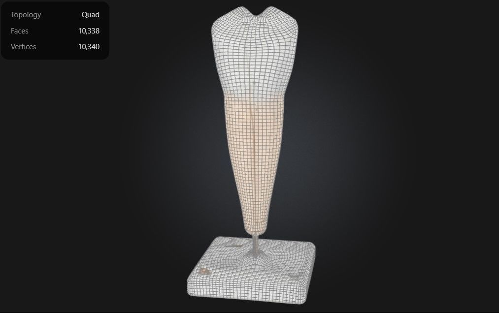
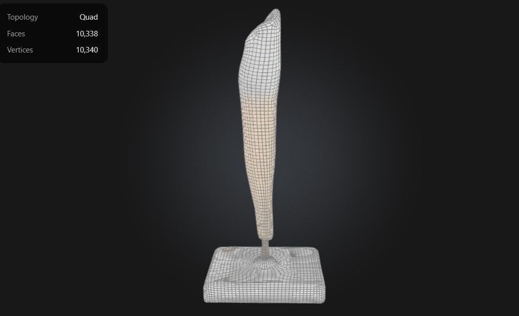
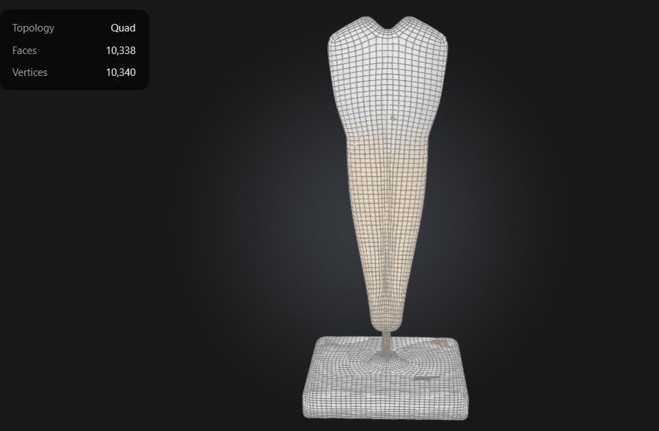
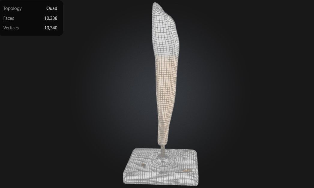
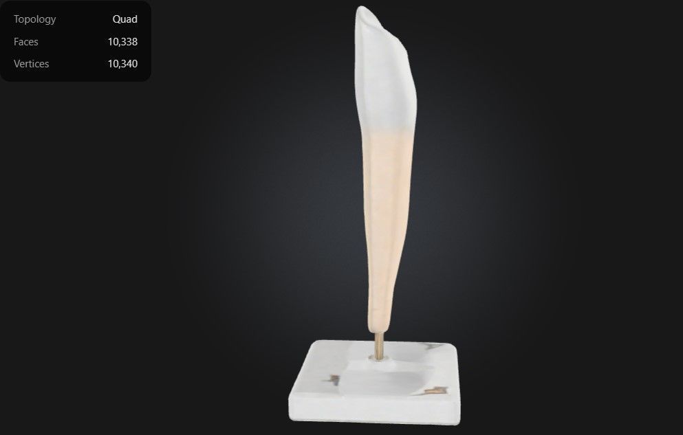
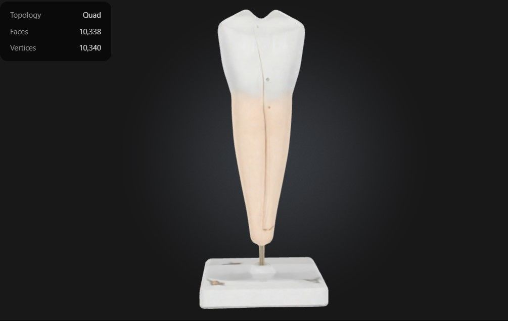
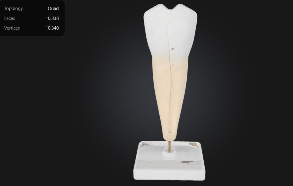
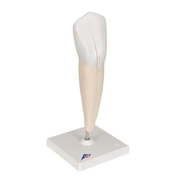
This 3D model of a lower canine tooth provides a highly detailed and anatomically accurate representation of the structure and function of a mandibular canine. It highlights key components, including the enamel, dentin, pulp chamber, and root canal, offering a clear visualization of the tooth’s internal and external anatomy. The model also showcases the sharp, pointed cusp designed for tearing and grasping food, along with its single, long root that provides strong anchorage and stability within the jawbone.
Designed for educational and clinical purposes, this model is an invaluable tool for studying dental anatomy, understanding root morphology, and visualizing conditions such as cavities, fractures, and periodontal diseases. Ideal for dentists, orthodontists, educators, and students, it supports teaching, training, and patient education by providing a hands-on approach to exploring the structure, function, and care of canine teeth.

3D Model formats
Format limitations
- Stereolithography (.stl)1010 KB
- Autodesk FBX (.fbx)4.99 MB
- PNG (.png) (2 files)7.56 MB
- glTF (.gltf, .glb)1.05 MB
- Blender (.blend)3.58 MB
- USDZ (.usdz)935 KB
- OBJ (.obj, .mtl)2.57 MB
3D Model details
- Publish date2025-01-05
- Model ID#5757803
- Ready for 3D Printing
Similar Models