
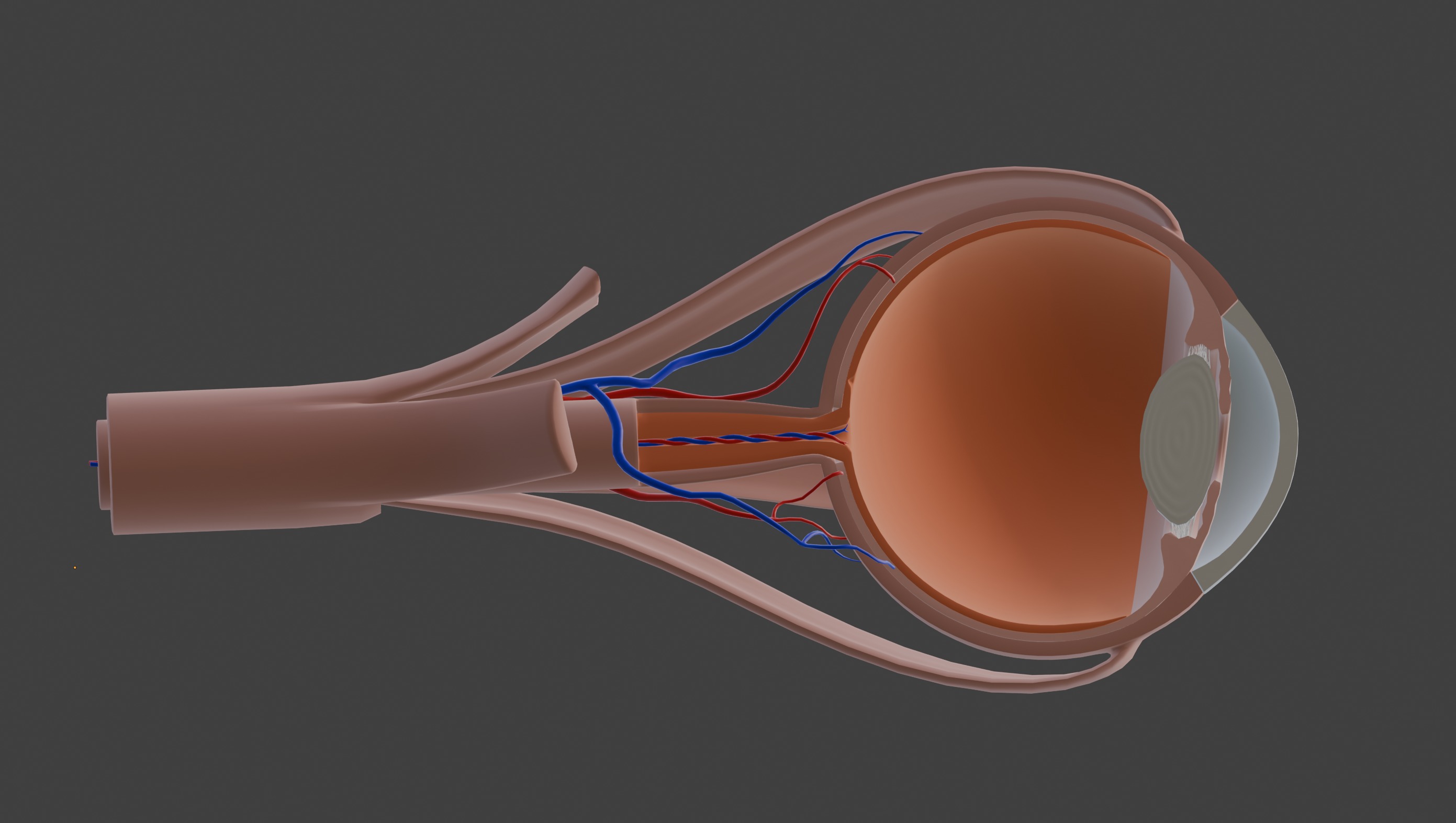


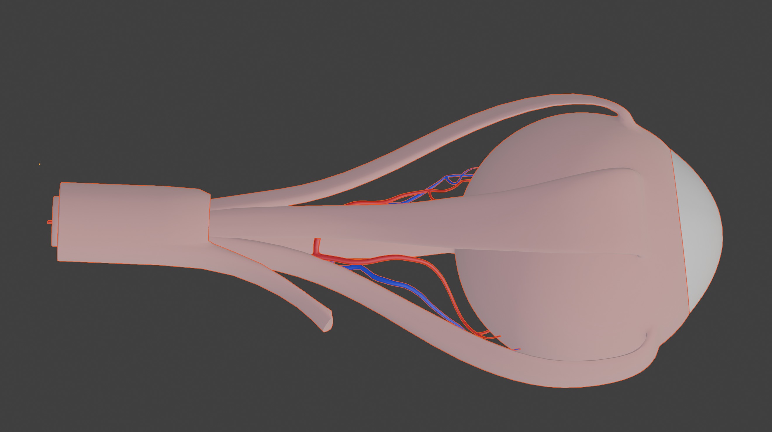


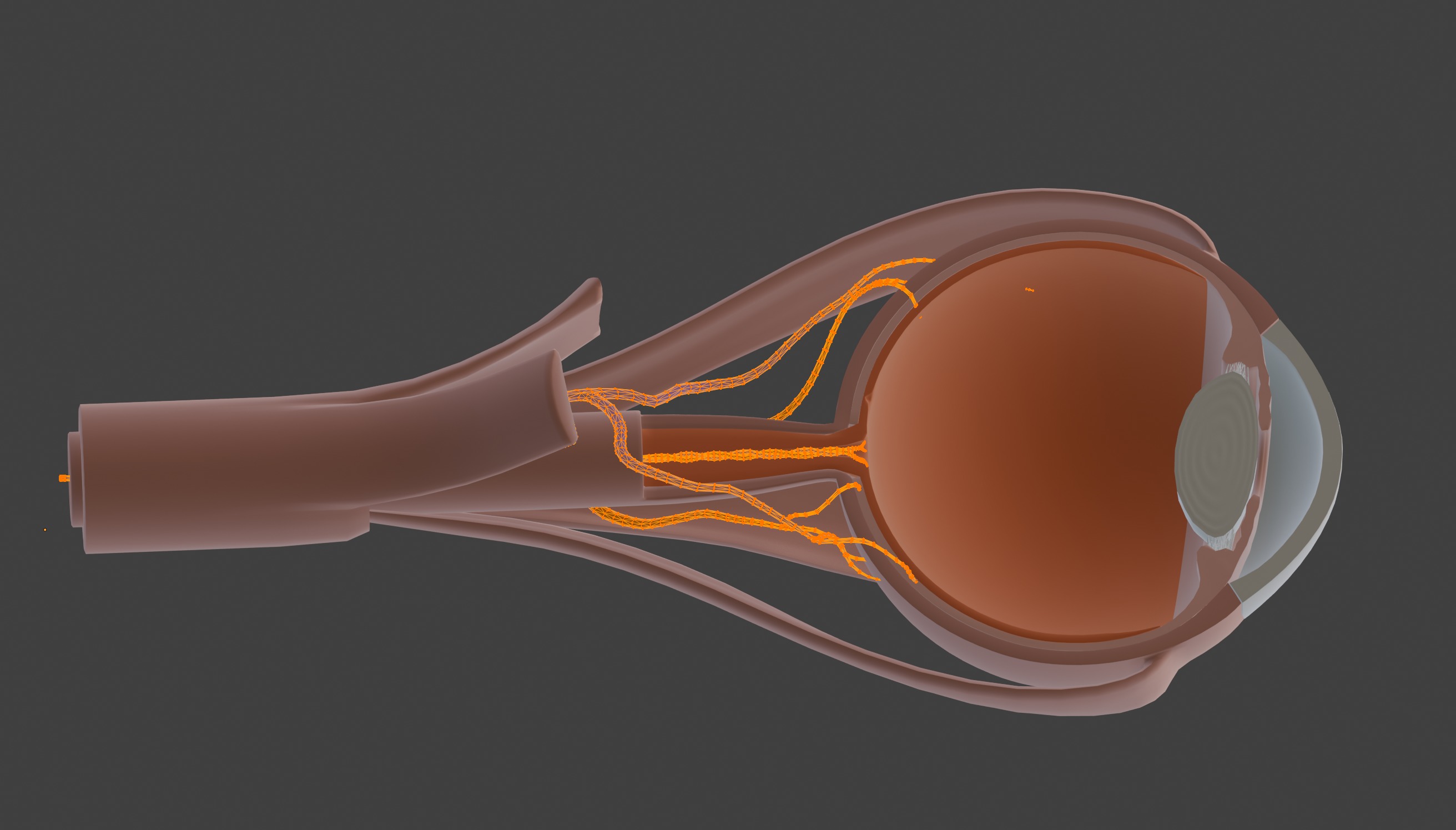
3D Model of Human Eye Anatomy 3D print model
This 3D model of root canal vs. filling provides a highly detailed and anatomically accurate comparison of two common dental treatments for tooth restoration. It highlights the internal structure of teeth, including the enamel, dentin, pulp chamber, and root canals, while illustrating the differences between a standard dental filling and a root canal procedure. The model clearly demonstrates how fillings are used to treat minor cavities by restoring damaged enamel, whereas root canal therapy is used to address deeper infections by cleaning, shaping, and sealing the root canals to preserve the tooth.
Designed for educational and clinical purposes, this model is an invaluable tool for studying dental anatomy, understanding treatment options, and visualizing conditions such as cavities, pulp infections, and root damage.
Ideal for dentists, endodontists, educators, and students, it supports teaching, training, and patient education by providing a hands-on approach to explaining dental procedures, helping patients make informed decisions about oral health care.
Included file formats: blend, dae, obj, stl,

3D Model formats
Format limitations
- Blender (.blend)15.2 MB
- Collada (.dae)11.7 MB
- Autodesk FBX (.fbx)3.13 MB
- OBJ (.obj, .mtl) (2 files)8.83 MB
- glTF (.gltf, .glb)2.67 MB
- Stereolithography (.stl)5.04 MB
3D Model details
- Ready for 3D Printing
- Publish date2025-01-07
- Model ID#5764413
Similar Models

Users who bought this item also bought...
