

3D Model of Anatomy Inside Human Eye 3D print model
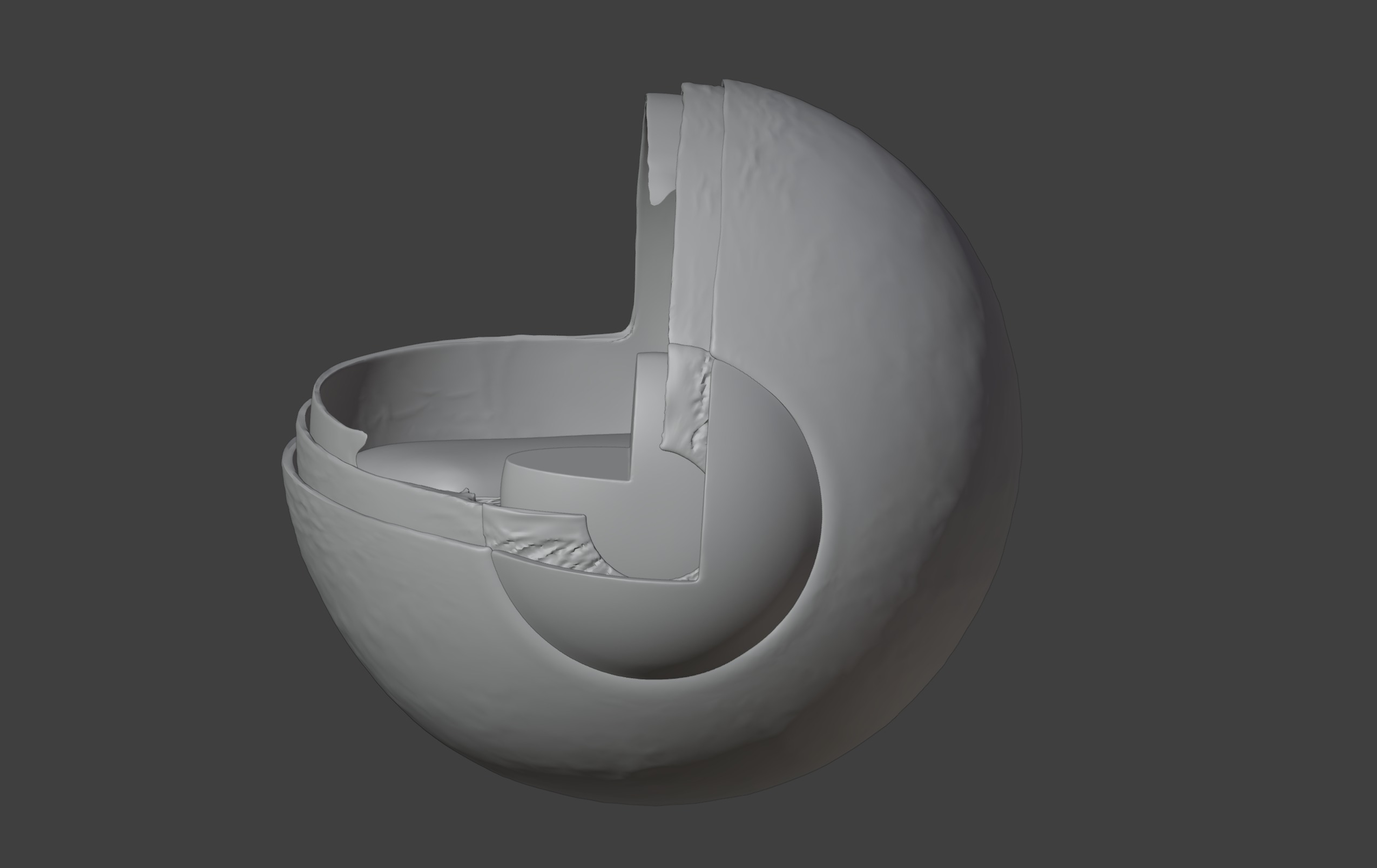
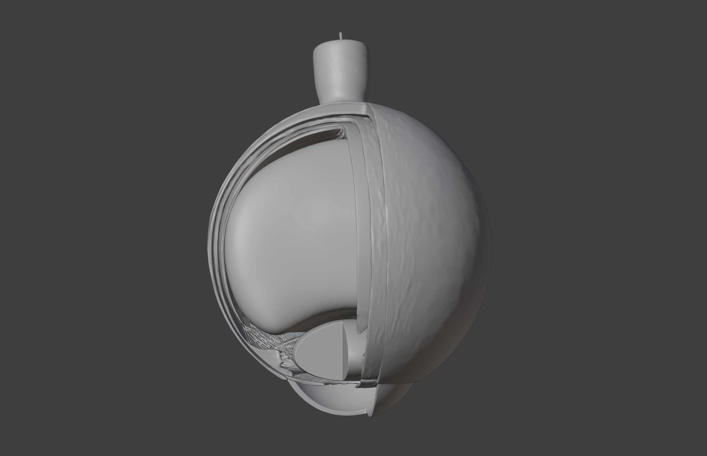
This 3D model of the anatomy inside the human eye provides a highly detailed and anatomically accurate representation of the internal structures of the eye. It highlights key components, including the cornea, iris, pupil, lens, retina, macula, optic nerve, and vitreous body, offering a clear visualization of how light is focused and processed to create vision.
The model also illustrates essential layers, such as the sclera, choroid, and retinal pigment epithelium, demonstrating their roles in protecting and nourishing the eye.
Designed for educational and clinical purposes, this model is an invaluable tool for studying ocular anatomy, understanding visual pathways, and exploring conditions such as cataracts, glaucoma, macular degeneration, and retinal detachment.
Ideal for ophthalmologists, optometrists, educators, and students, it supports teaching, training, and patient education by providing a hands-on approach to learning about the structure, function, and disorders of the human eye.
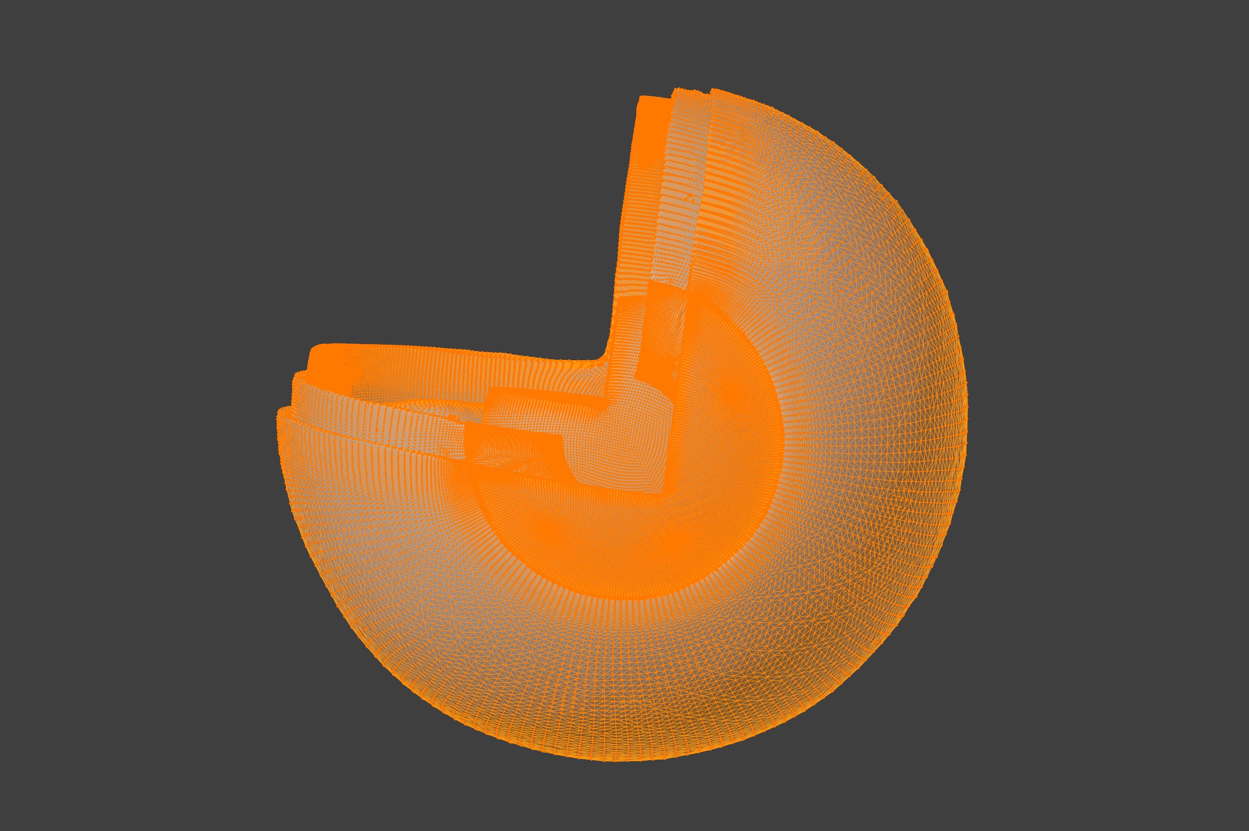
Included file formats: blend, dae, fbx, obj, stl

3D Model formats
Format limitations
- Collada (.dae)120 MB
- OBJ (.obj, .mtl) (2 files)81.3 MB
- Blender (.blend)72.7 MB
- glTF (.gltf, .glb)21.6 MB
- Stereolithography (.stl)38.9 MB
3D Model details
- Ready for 3D Printing
- Publish date2025-01-07
- Model ID#5764583
Similar Models

Users who bought this item also bought...
