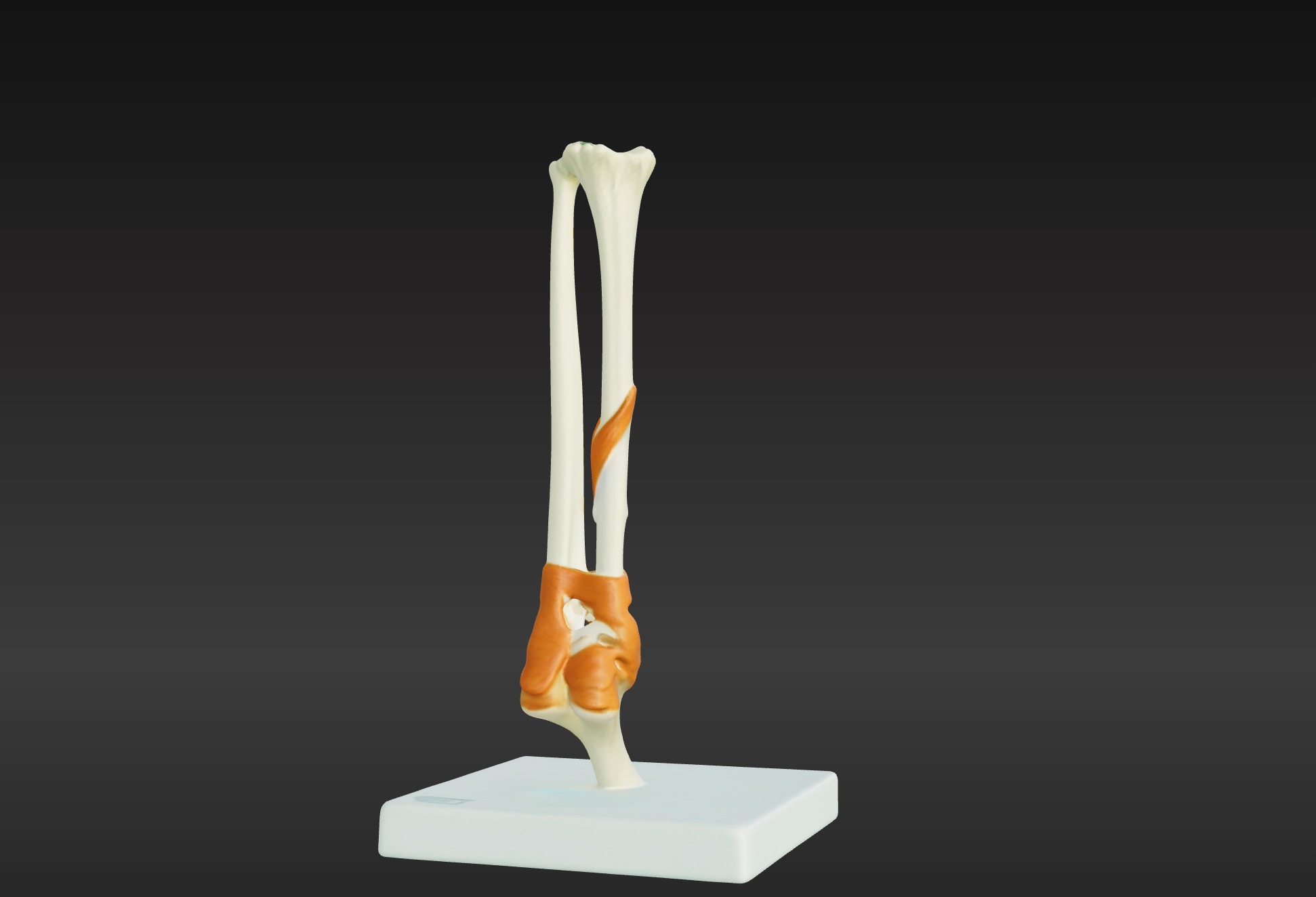
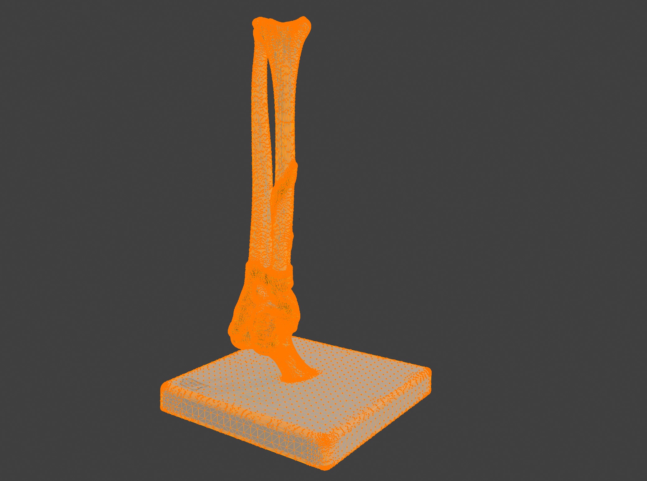
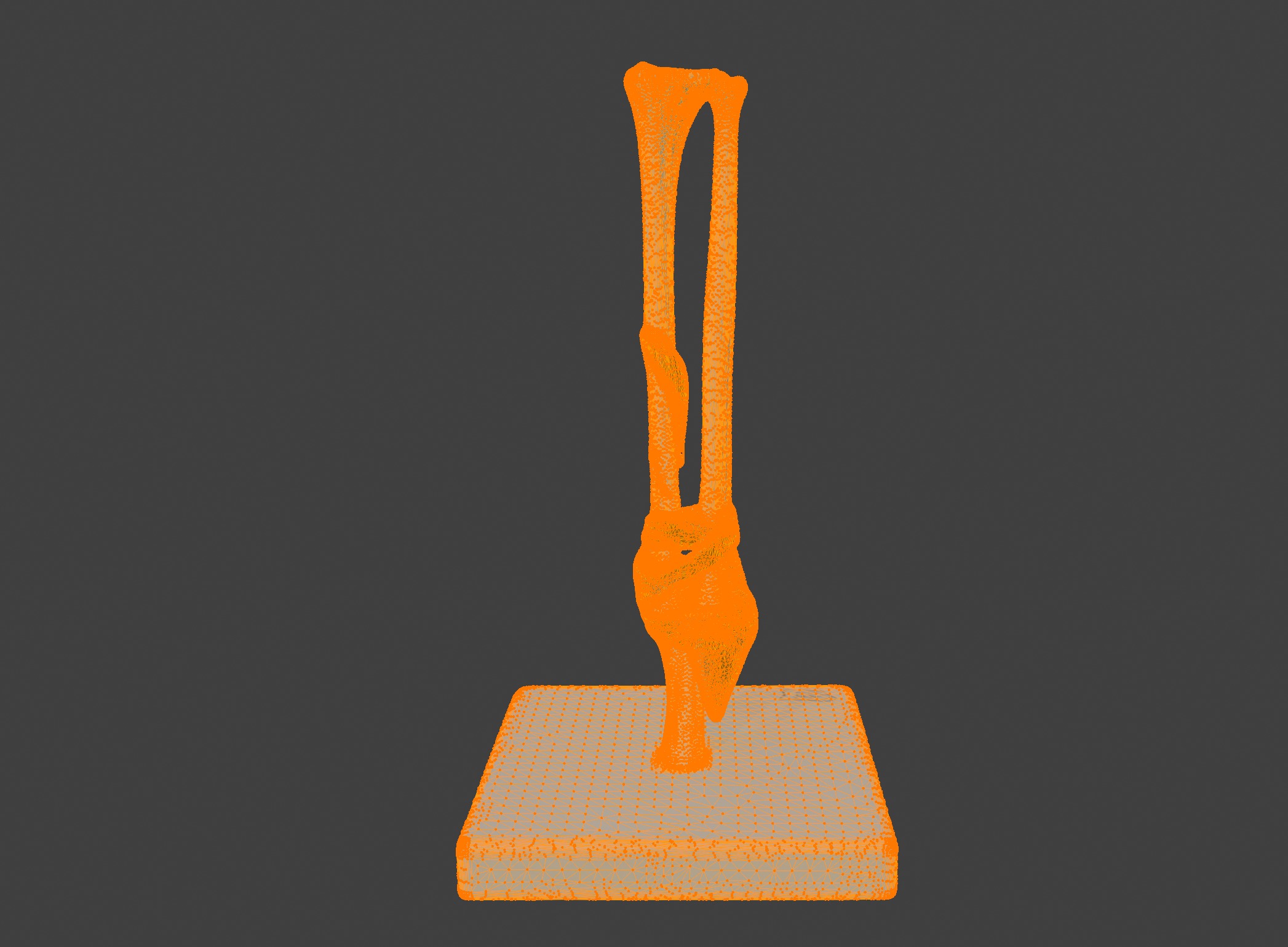
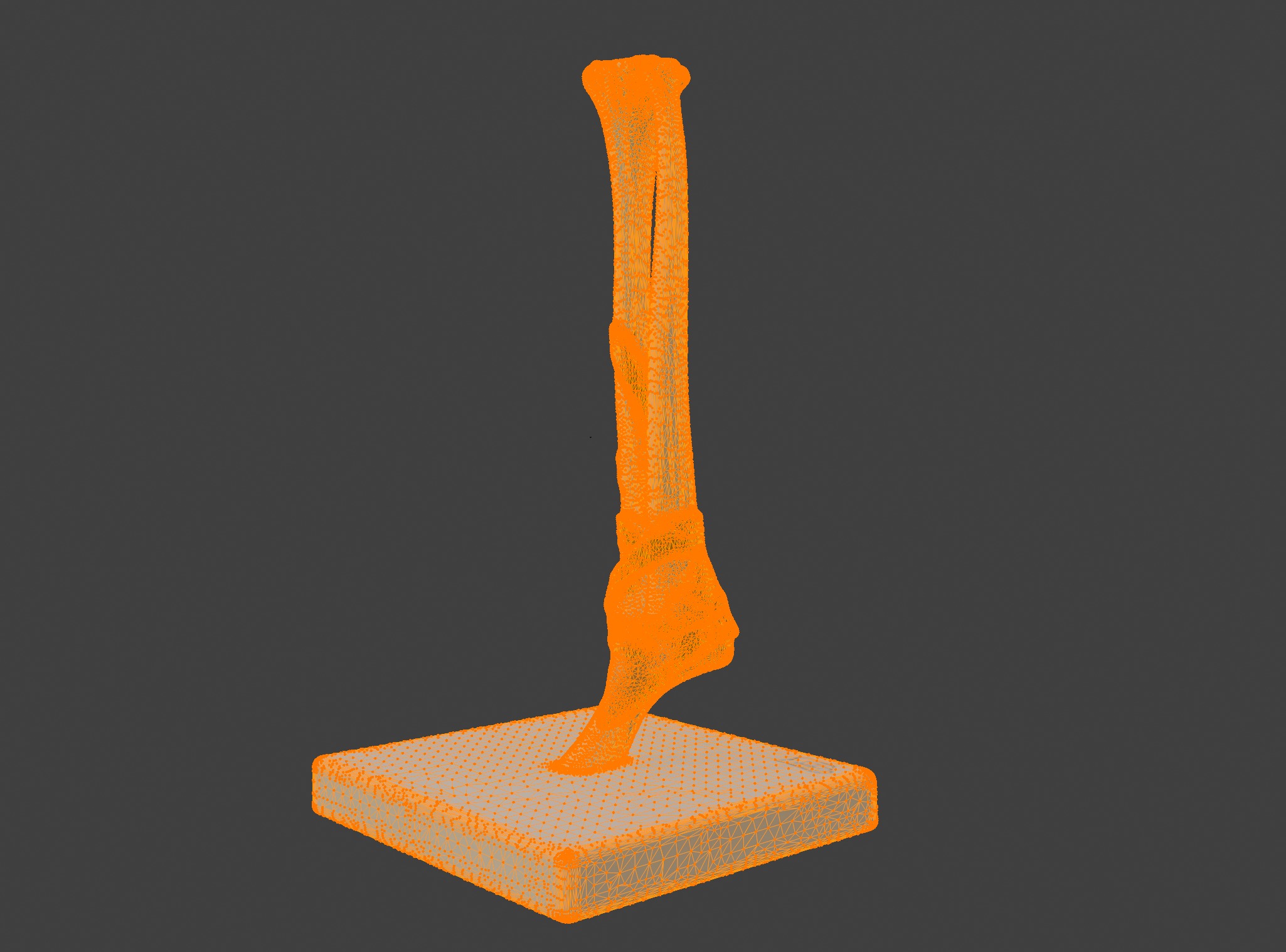
3D Joint Anatomy Model with Stand 3D print model
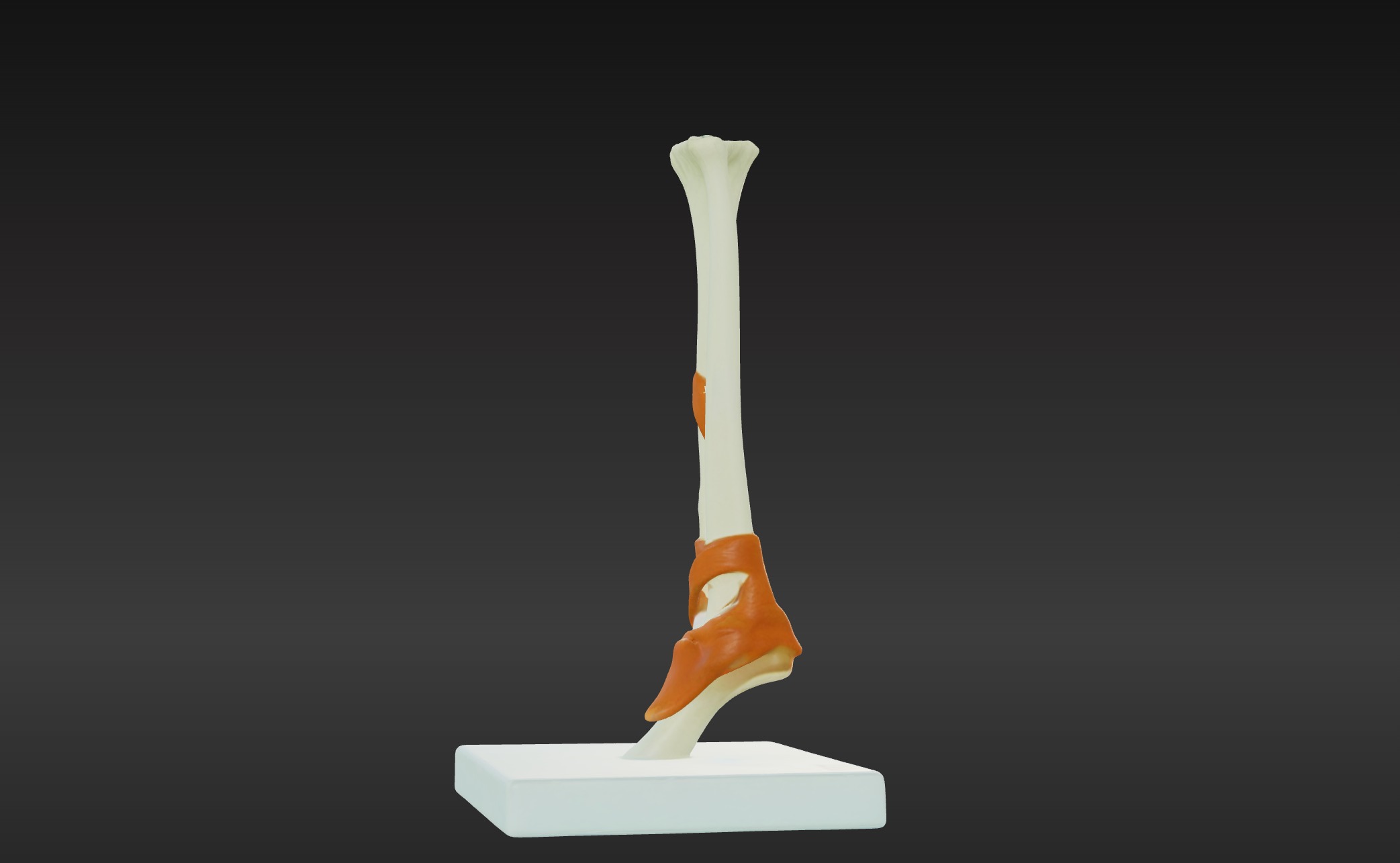
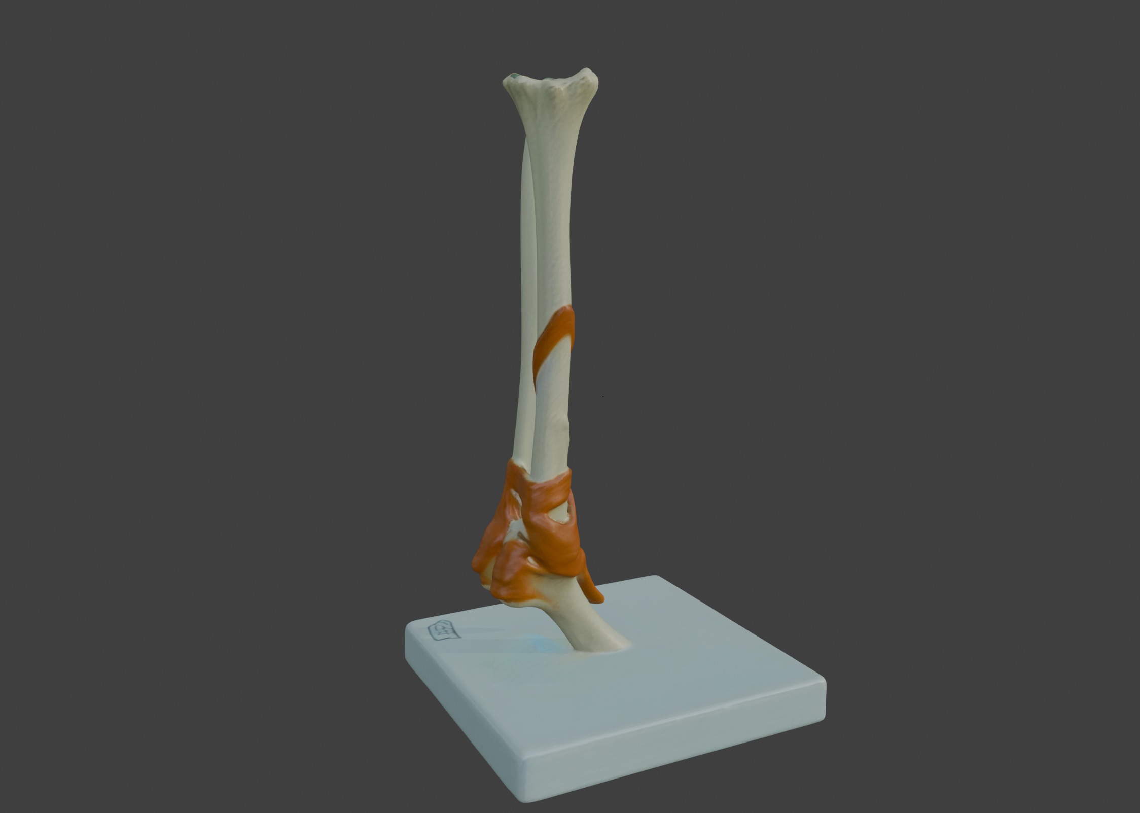
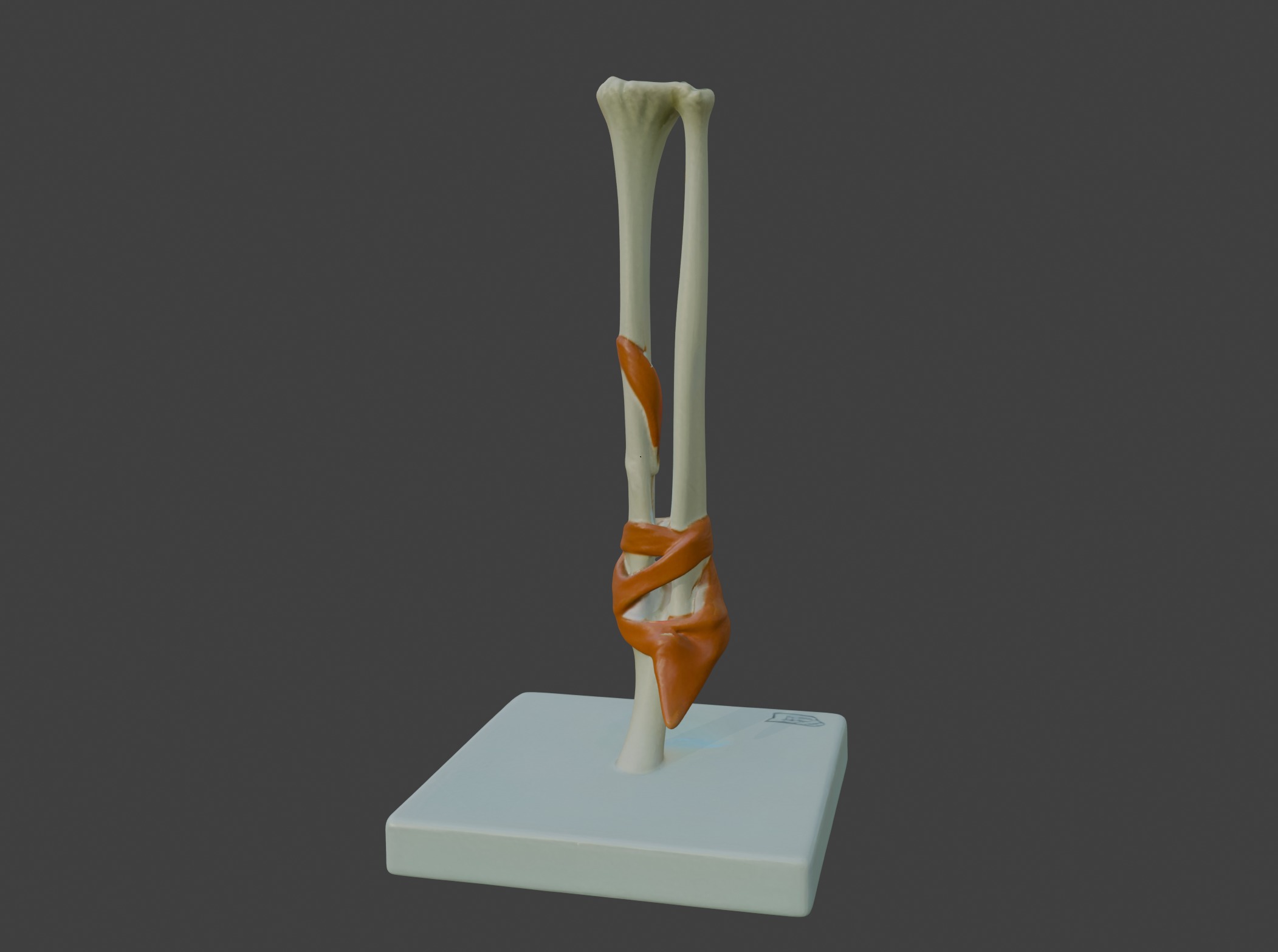
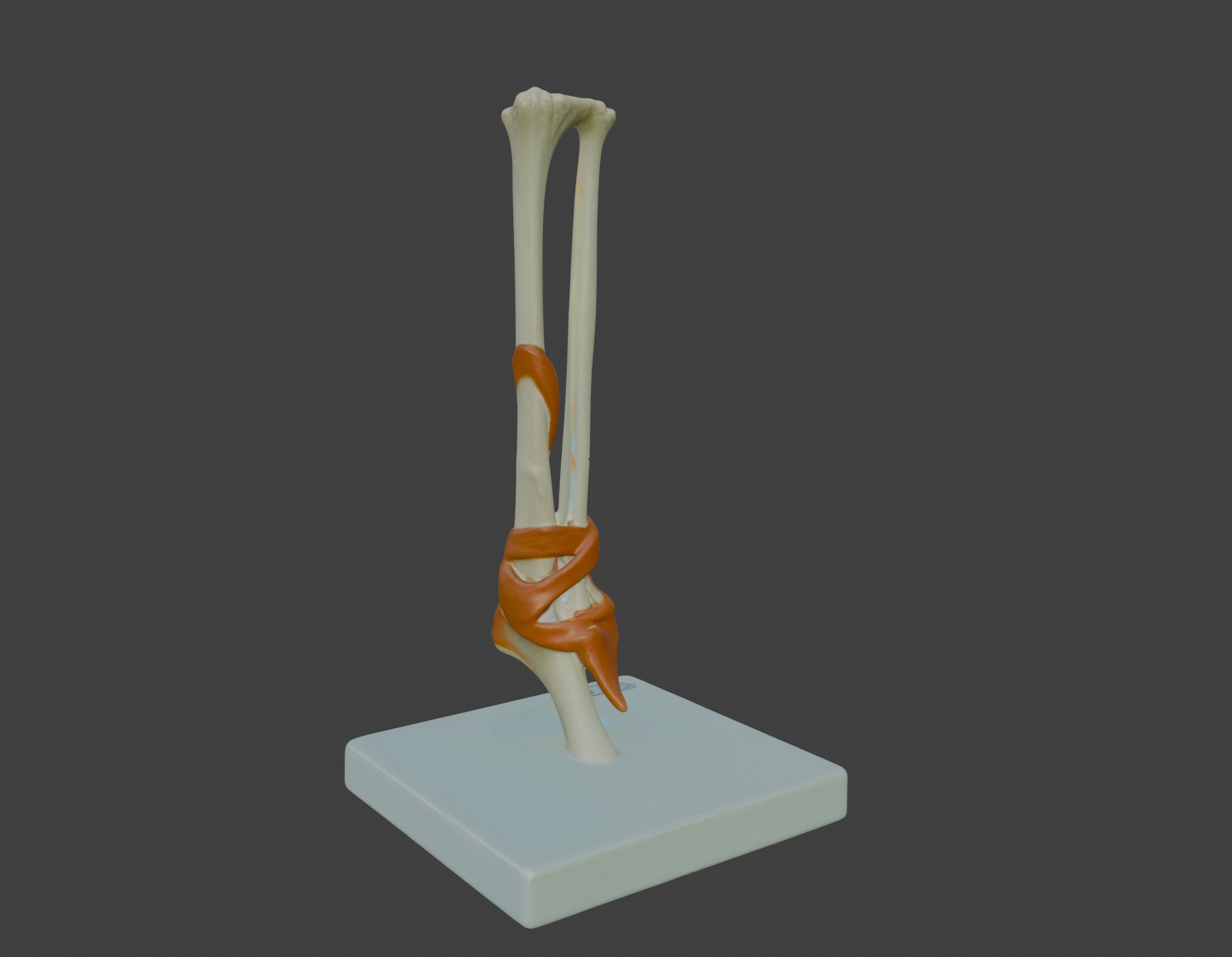
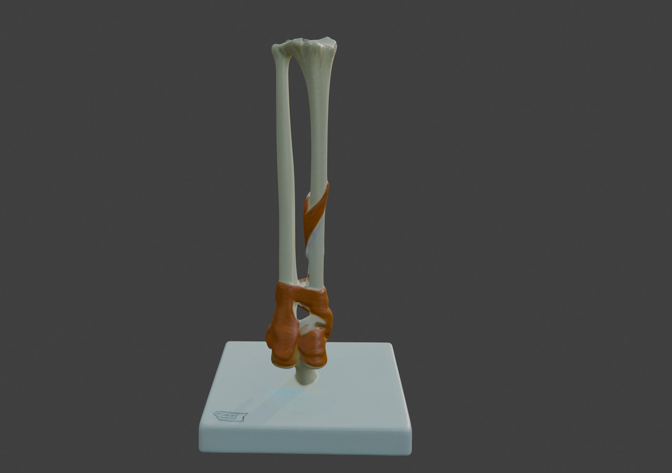
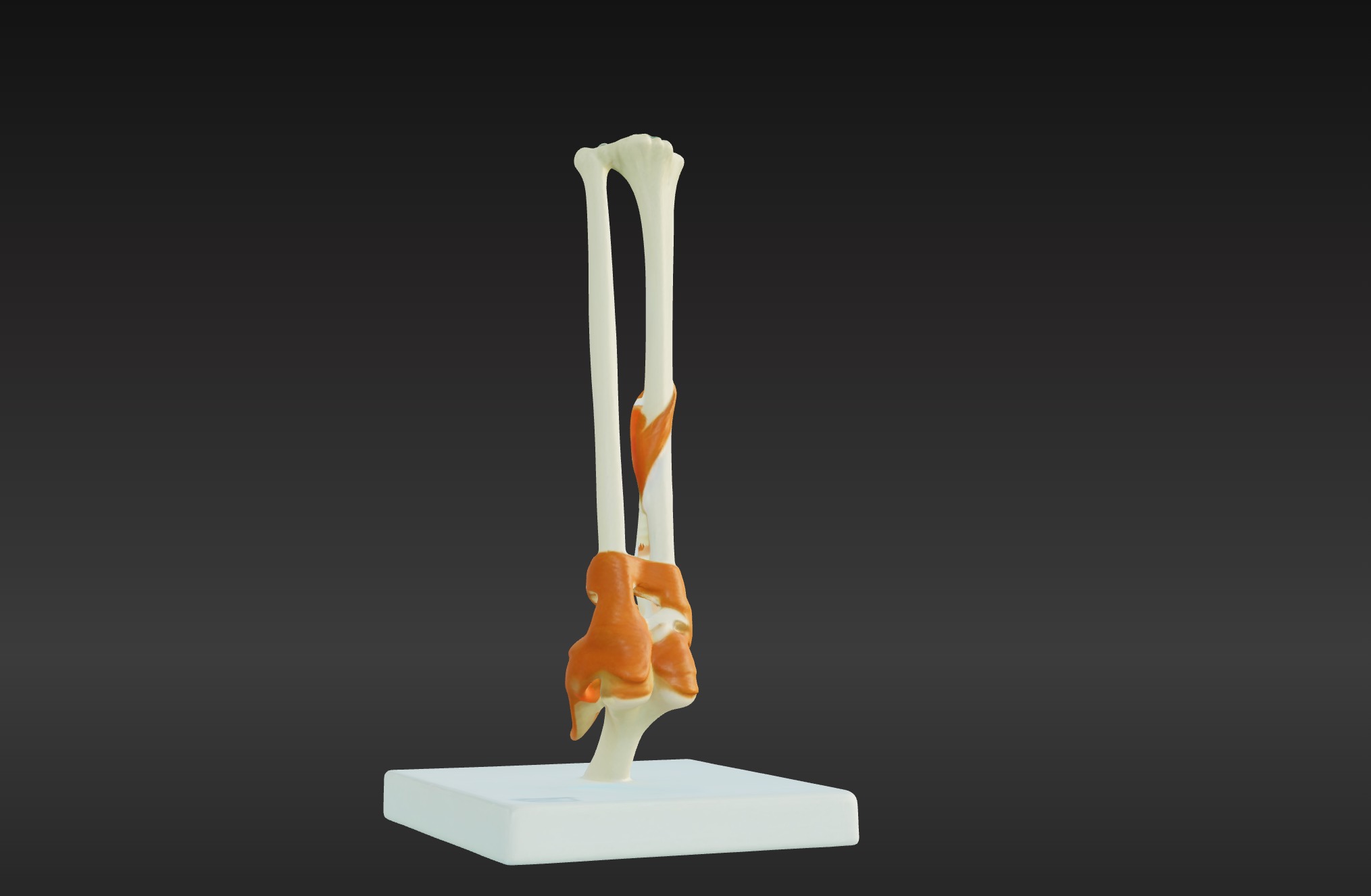
This 3D joint anatomy model with stand provides a highly detailed and anatomically accurate representation of a human joint, showcasing its structural components and functional mechanics. It highlights key features such as articular cartilage, synovial membrane, ligaments, tendons, and the bones that form the joint.
The model effectively demonstrates how joints enable movement while providing stability and support to the skeletal system.
Mounted on a sturdy stand for easy viewing, this model is ideal for educational and clinical purposes. It is an invaluable tool for studying joint anatomy, understanding biomechanics, and visualizing conditions such as arthritis, ligament injuries, and joint degeneration.
Perfect for orthopedic specialists, physical therapists, educators, and students, it supports teaching, training, and patient education by providing a hands-on approach to exploring the structure and function of human joints.

3D Model formats
Format limitations
- Blender (.blend)61.7 MB
- glTF (.gltf, .glb)3.53 MB
- Autodesk FBX (.fbx)2.53 MB
- OBJ (.obj, .mtl) (2 files)7.02 MB
- Collada (.dae)11 MB
- Stereolithography (.stl)3.63 MB
3D Model details
- Publish date2025-01-15
- Model ID#5786584
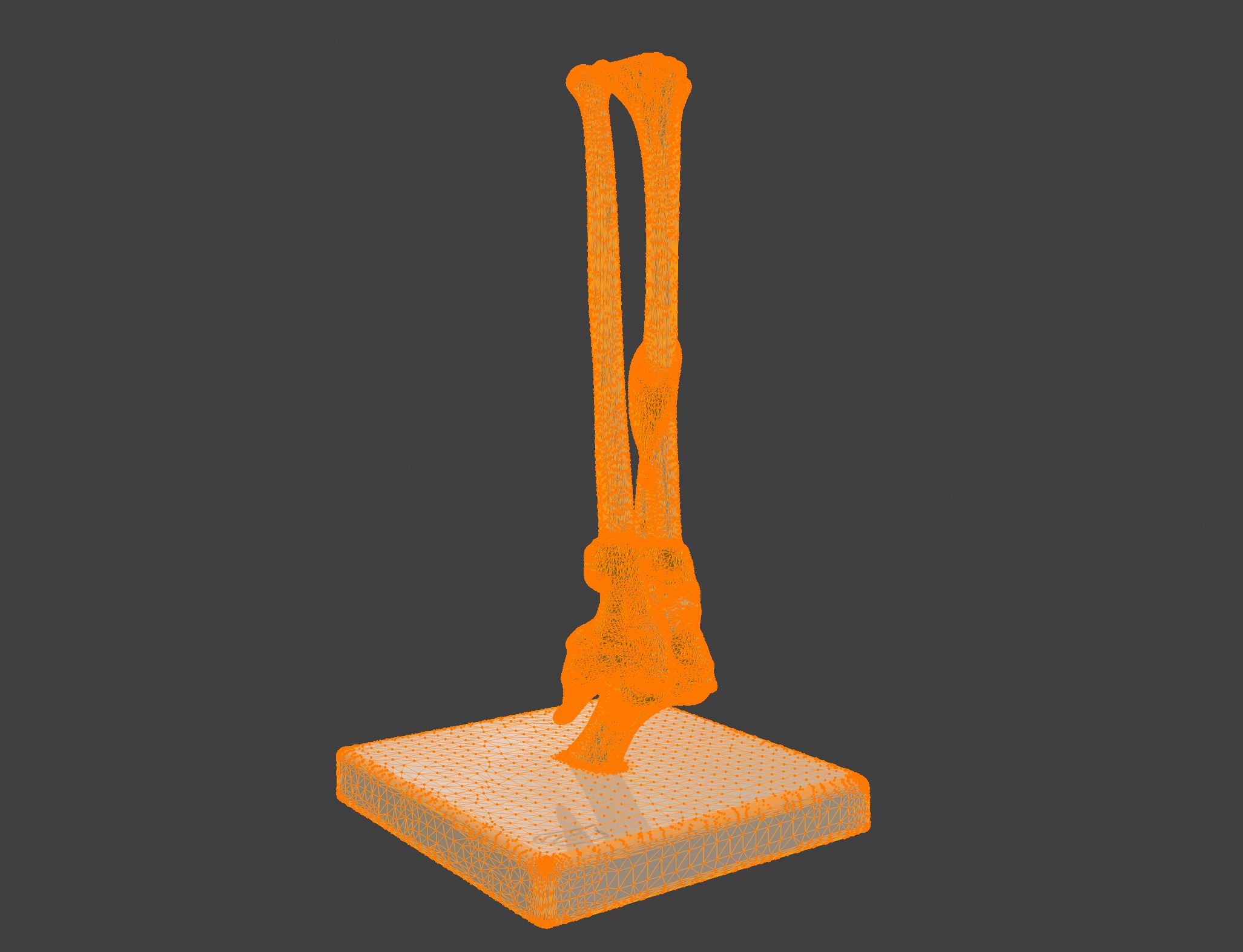
- Ready for 3D Printing
Similar Models