

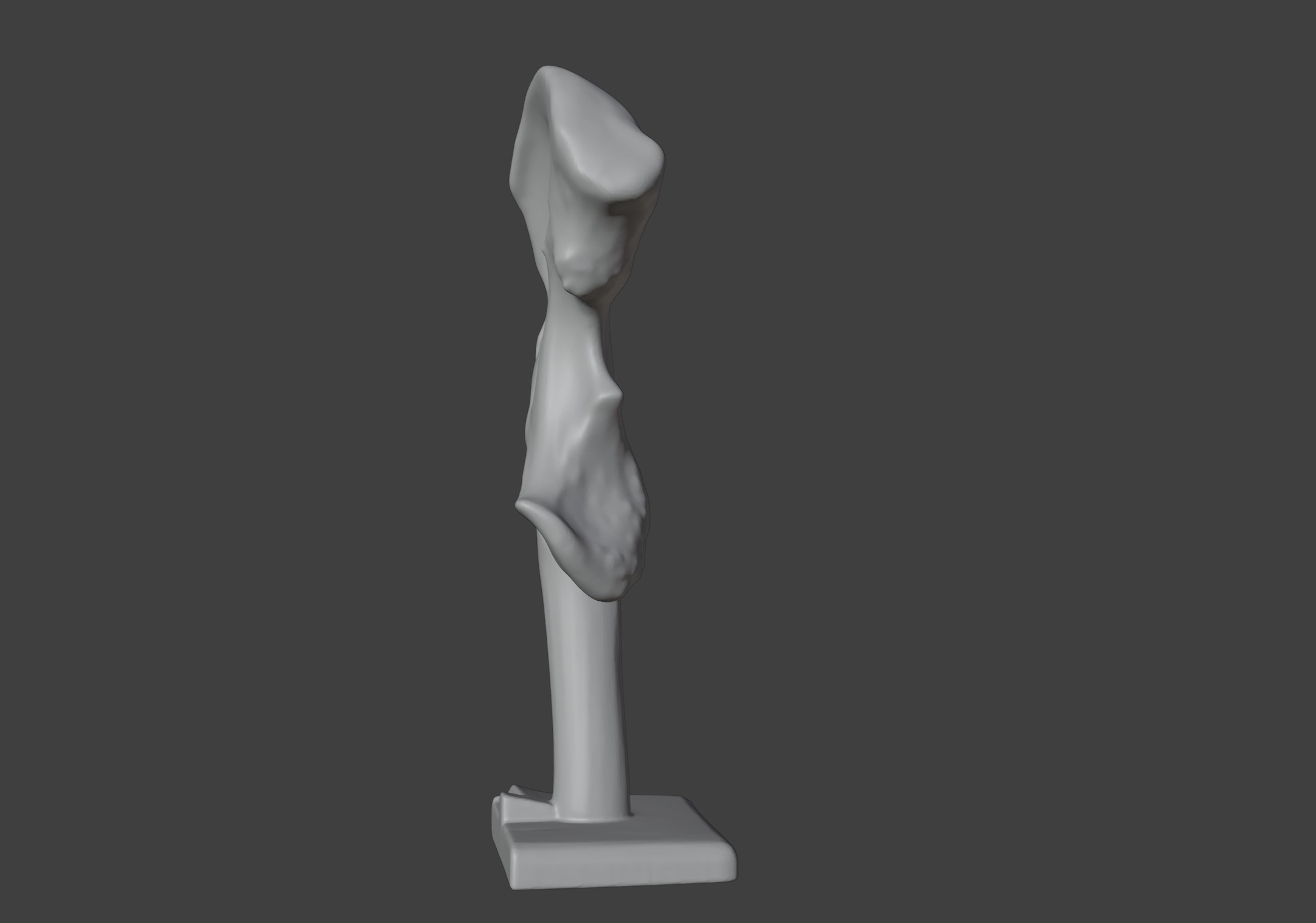
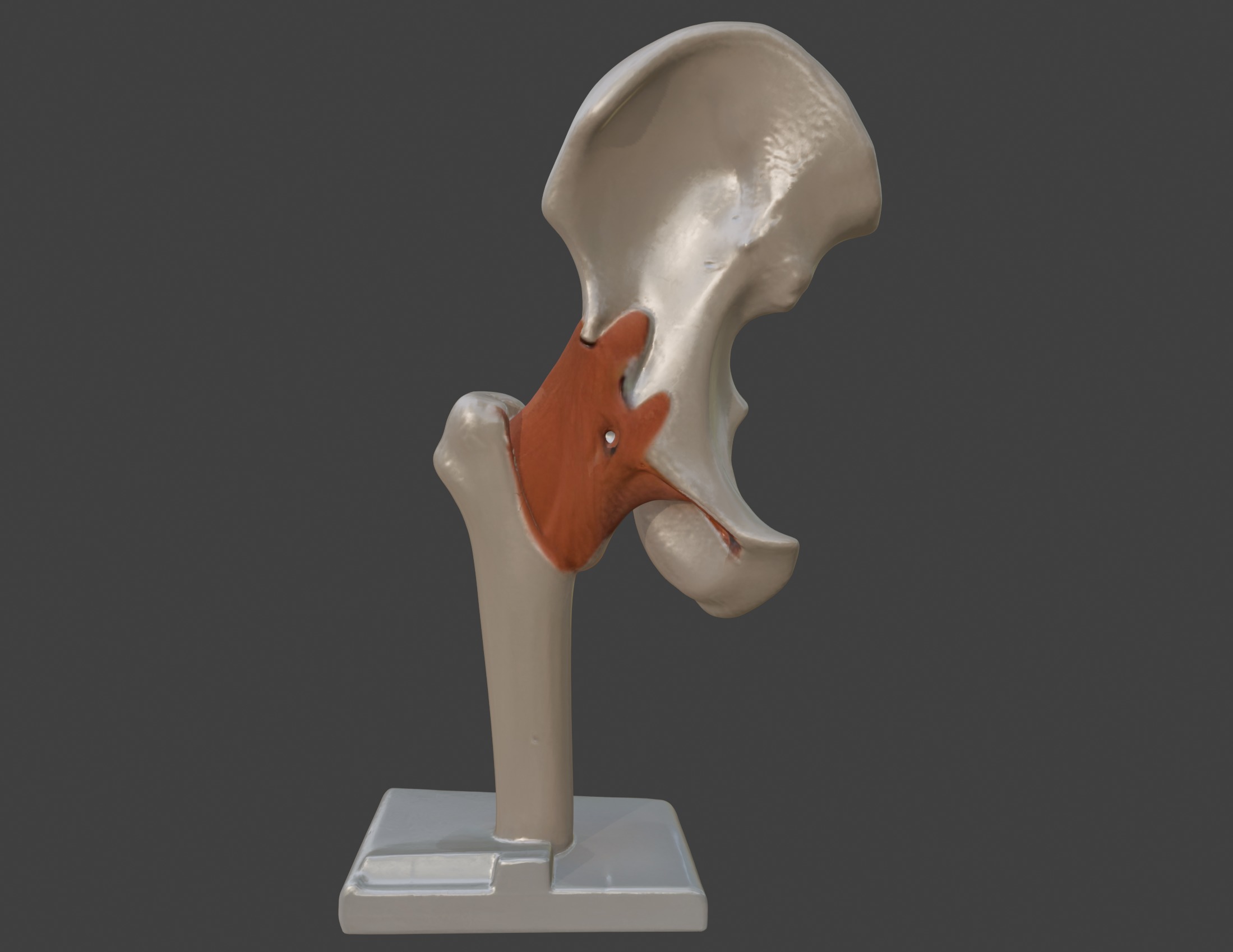
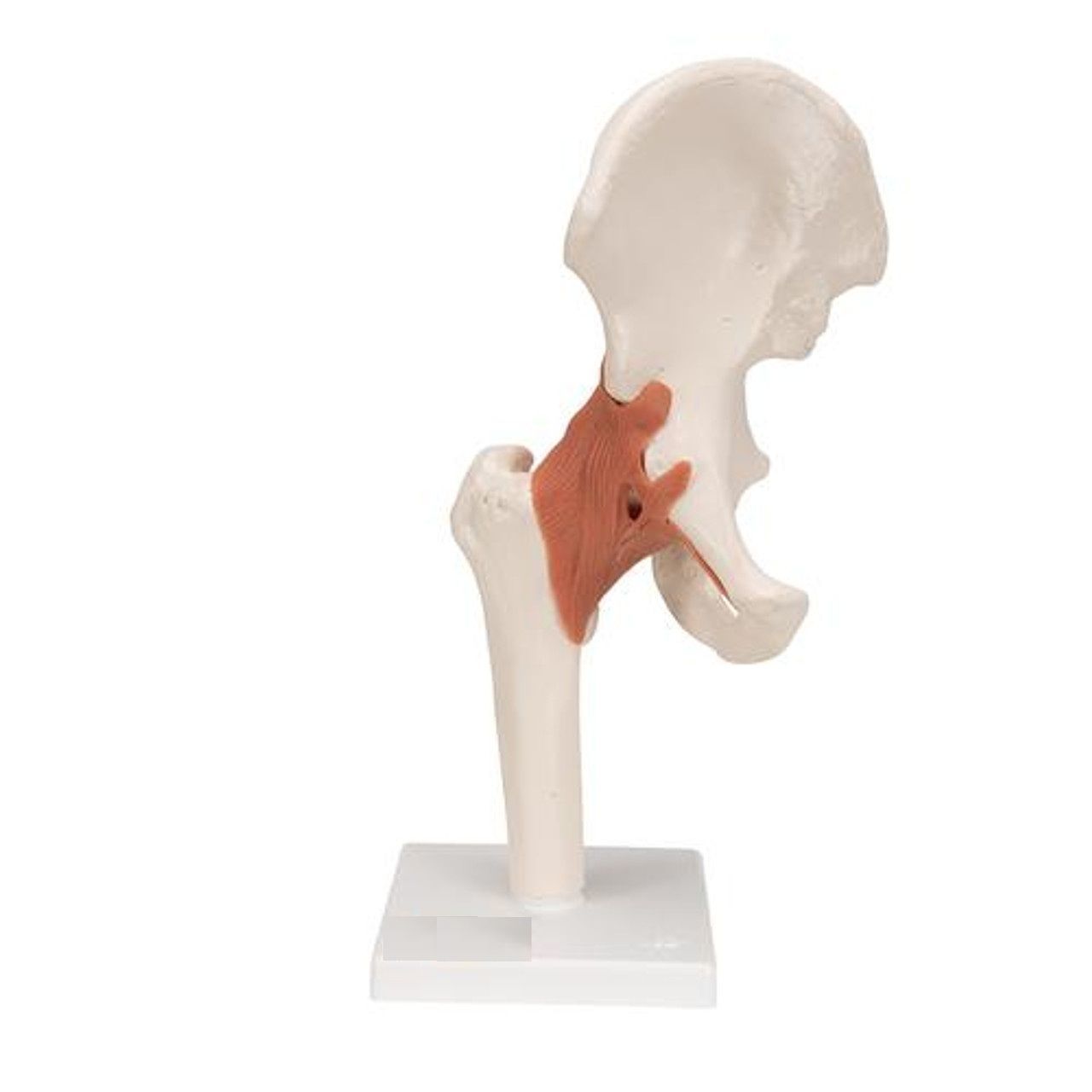


3D Hip Joint Anatomy Model 3D print model
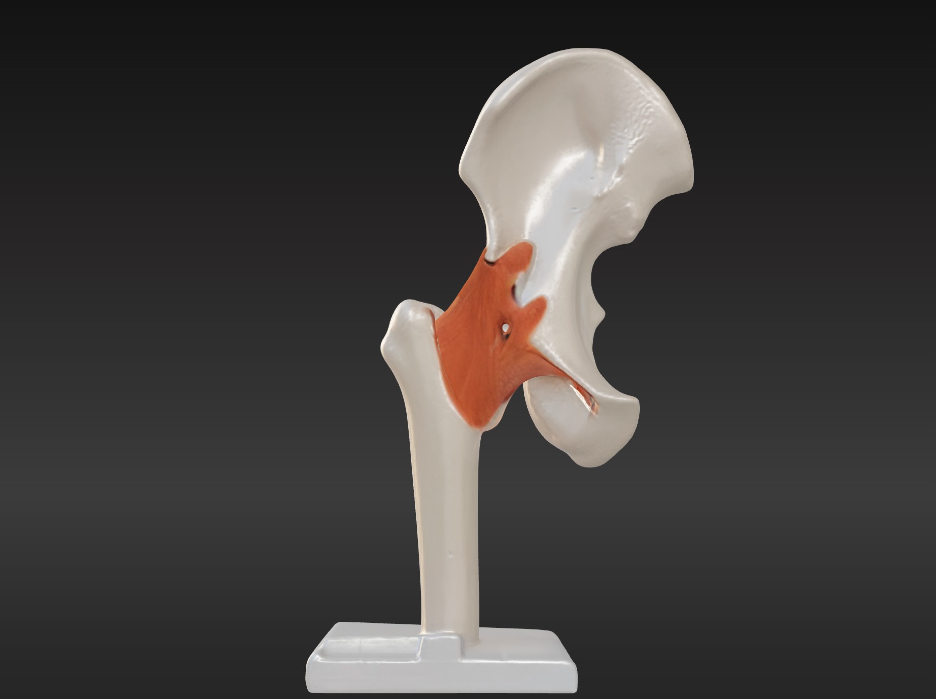
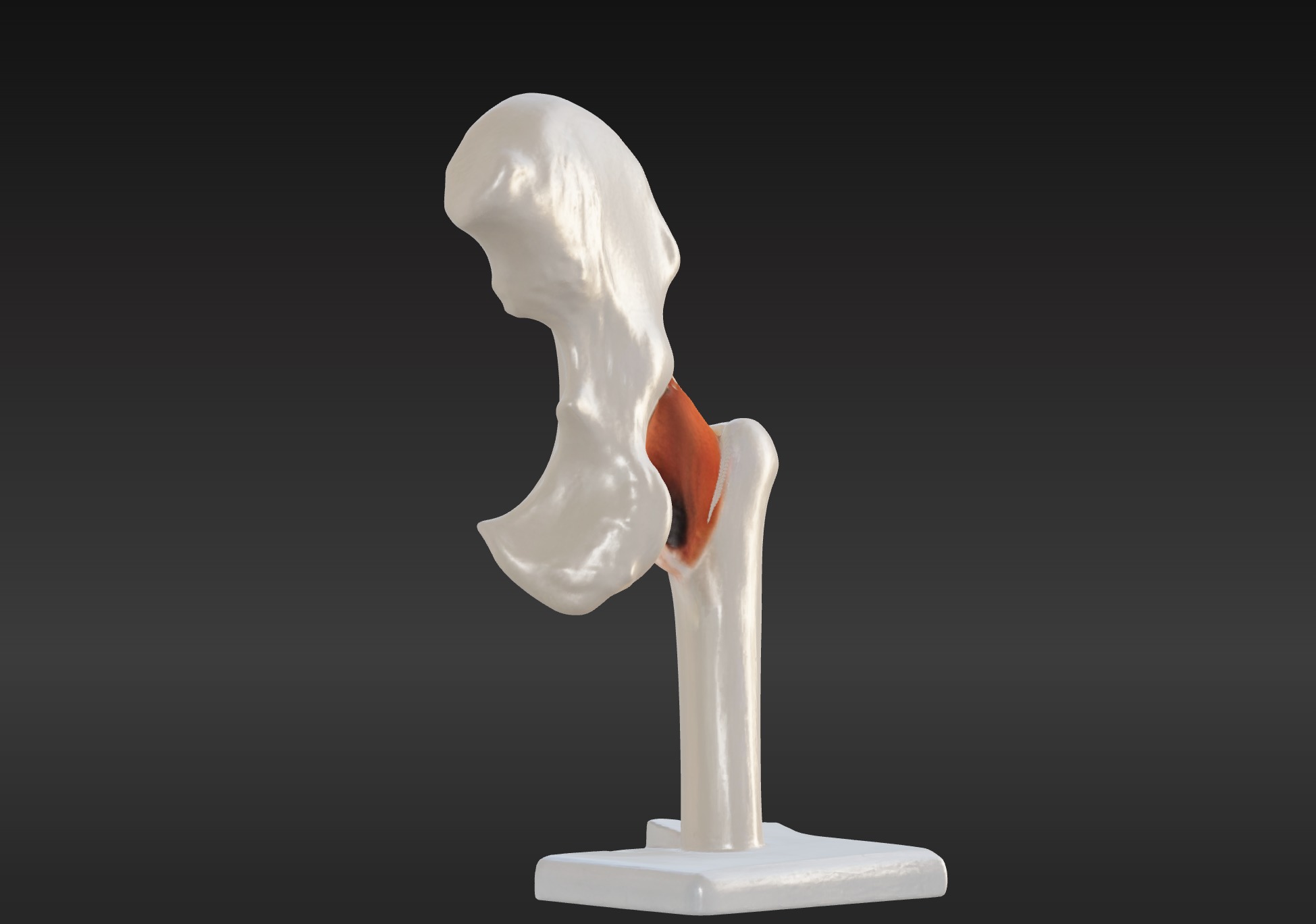
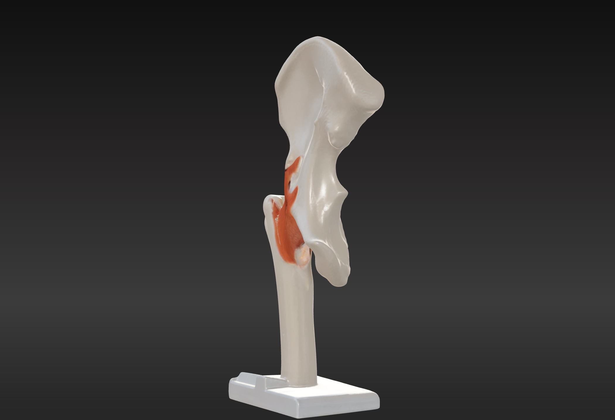
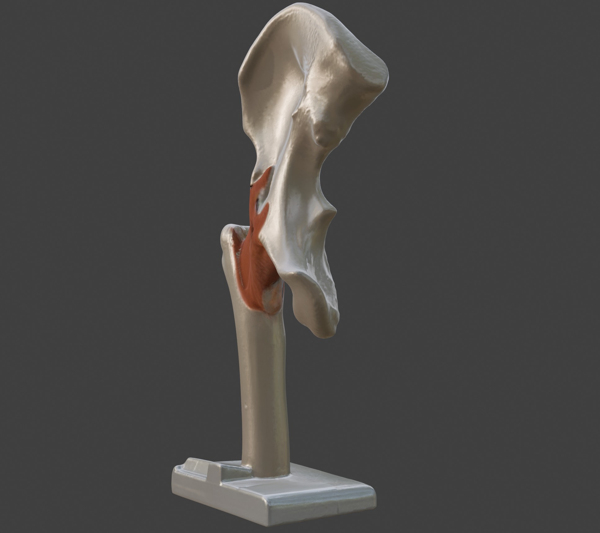
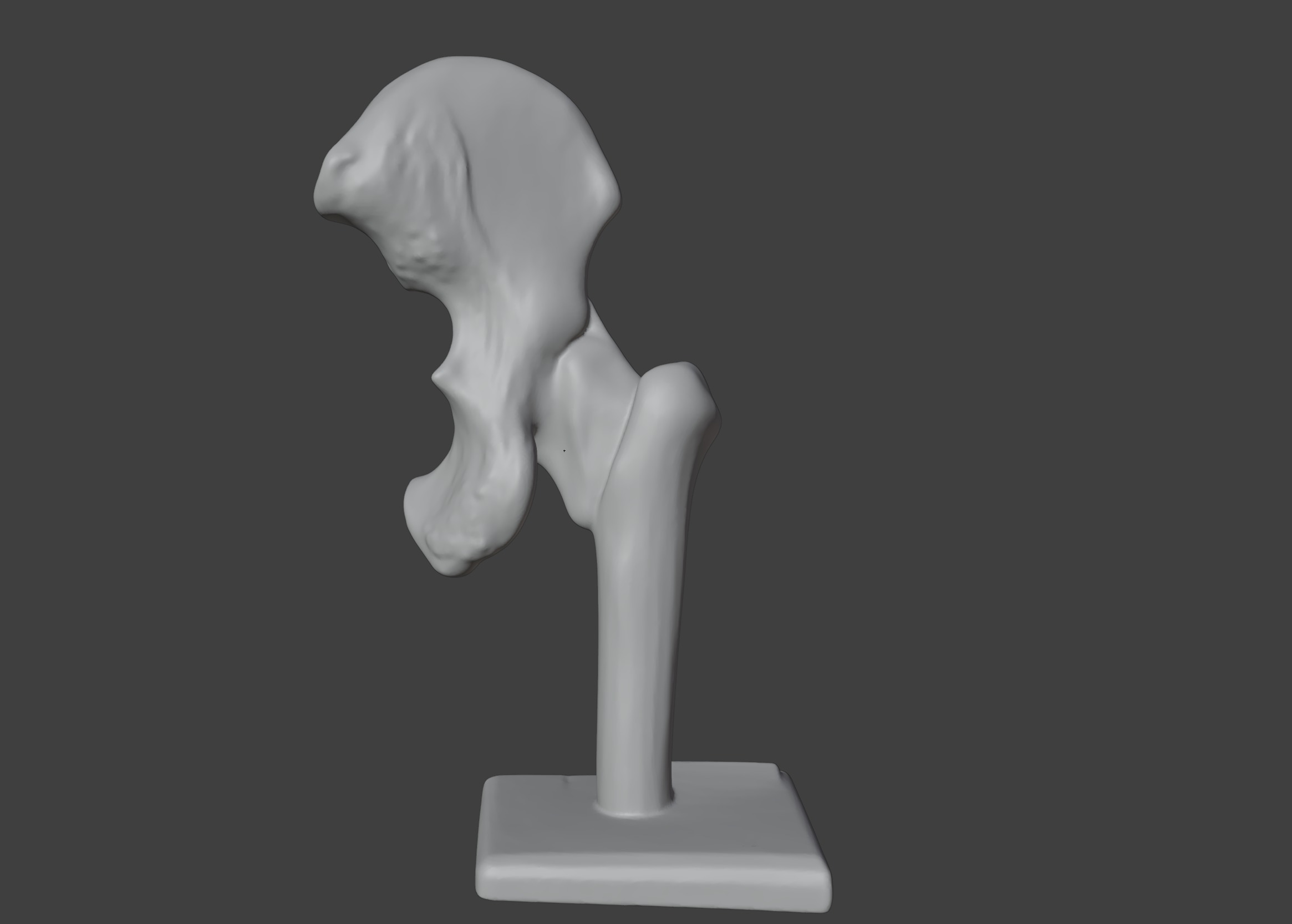
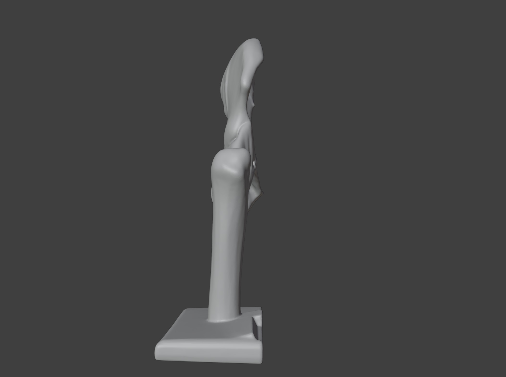
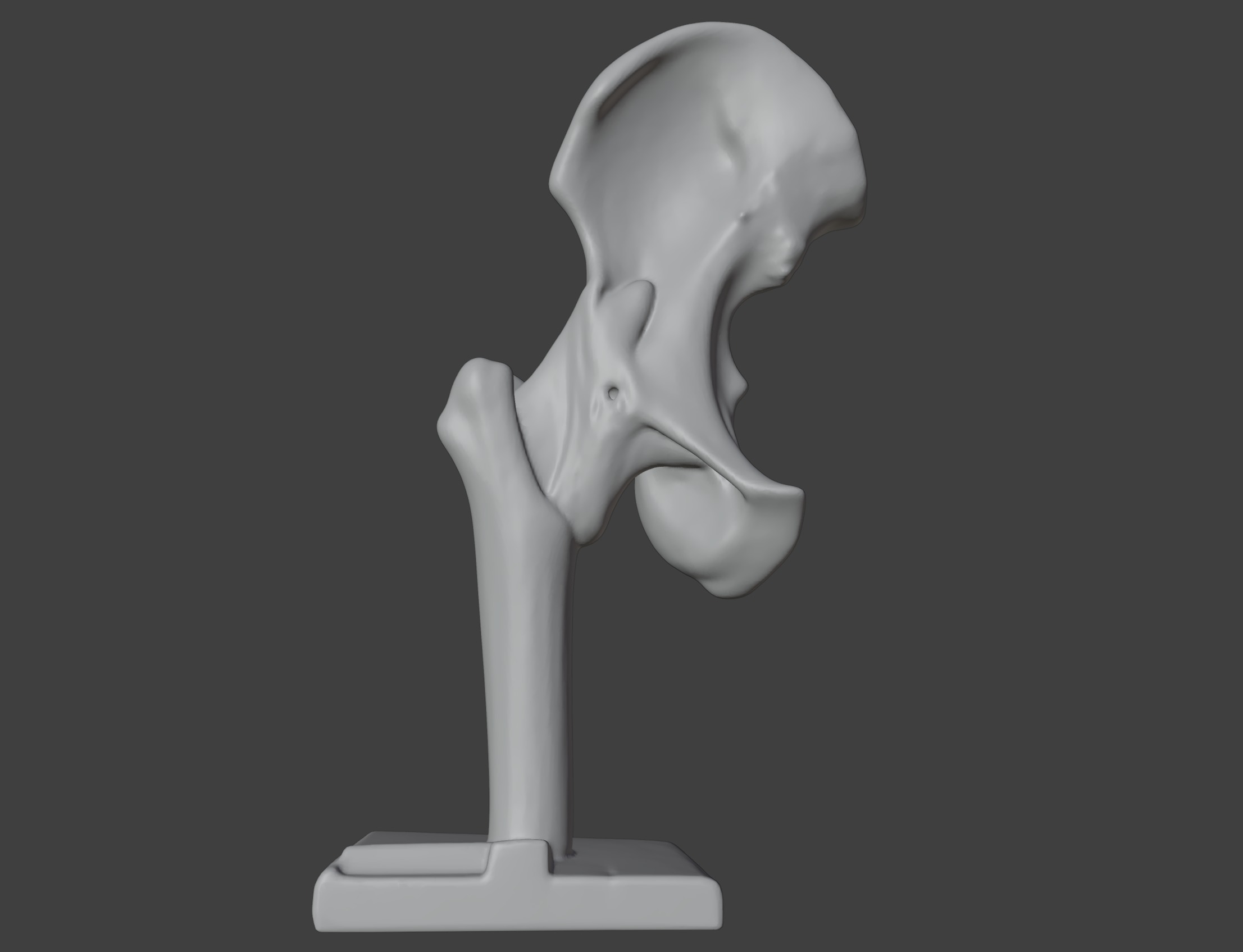
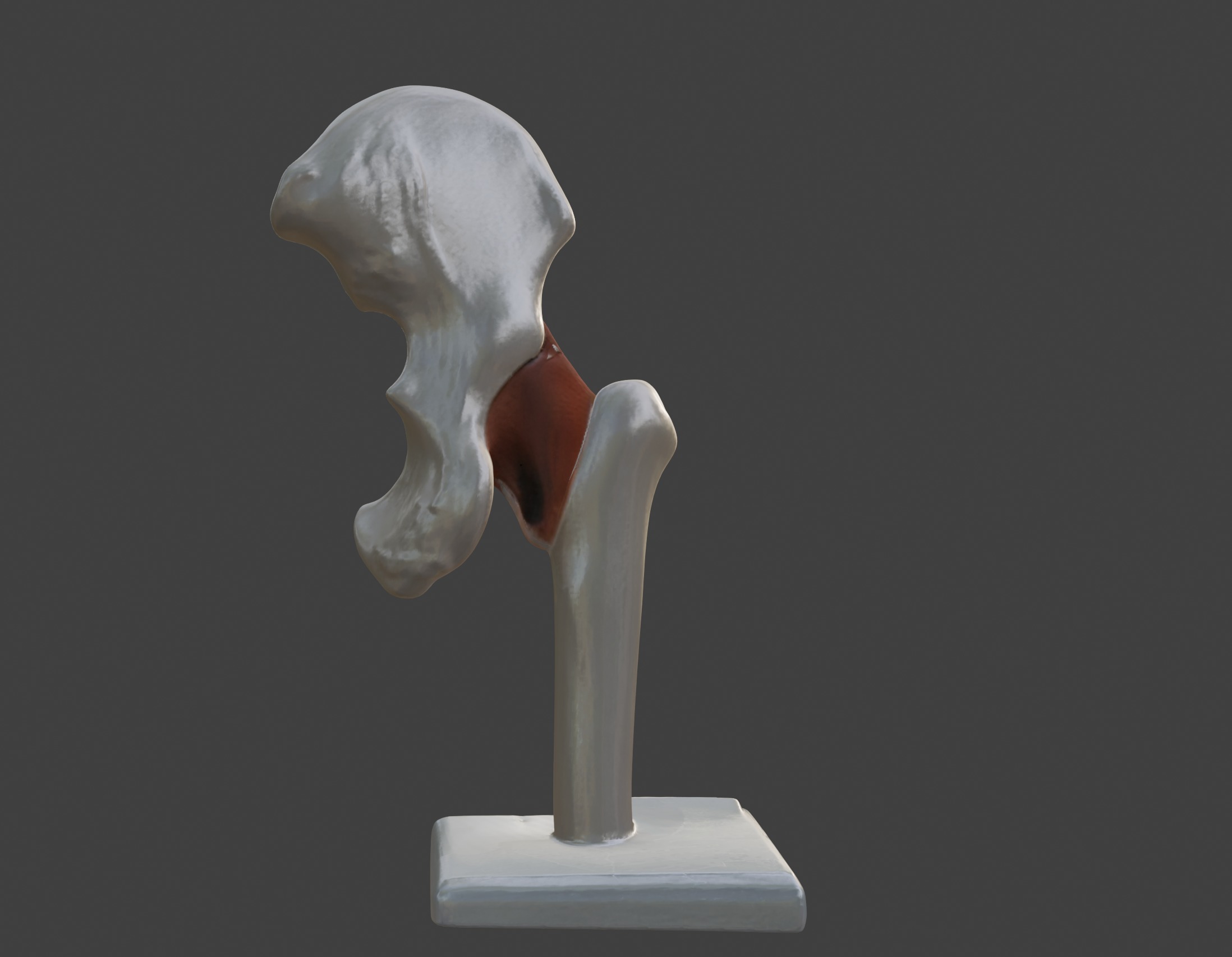
This 3D hip joint anatomy model provides a highly detailed and anatomically accurate representation of the human hip joint and its surrounding structures. It highlights key components, including the femoral head, acetabulum, articular cartilage, ligaments (such as the iliofemoral ligament), and surrounding muscles and tendons.
The model also demonstrates the ball-and-socket structure of the hip joint, offering insights into its range of motion and weight-bearing capabilities.
Designed for educational and clinical purposes, this model is an invaluable tool for studying musculoskeletal anatomy, understanding joint mechanics, and visualizing conditions such as arthritis, hip fractures, and labral tears.
Ideal for orthopedic specialists, physical therapists, educators, and students, it supports teaching, training, and patient education by providing a hands-on approach to exploring the structure, function, and health of the hip joint.

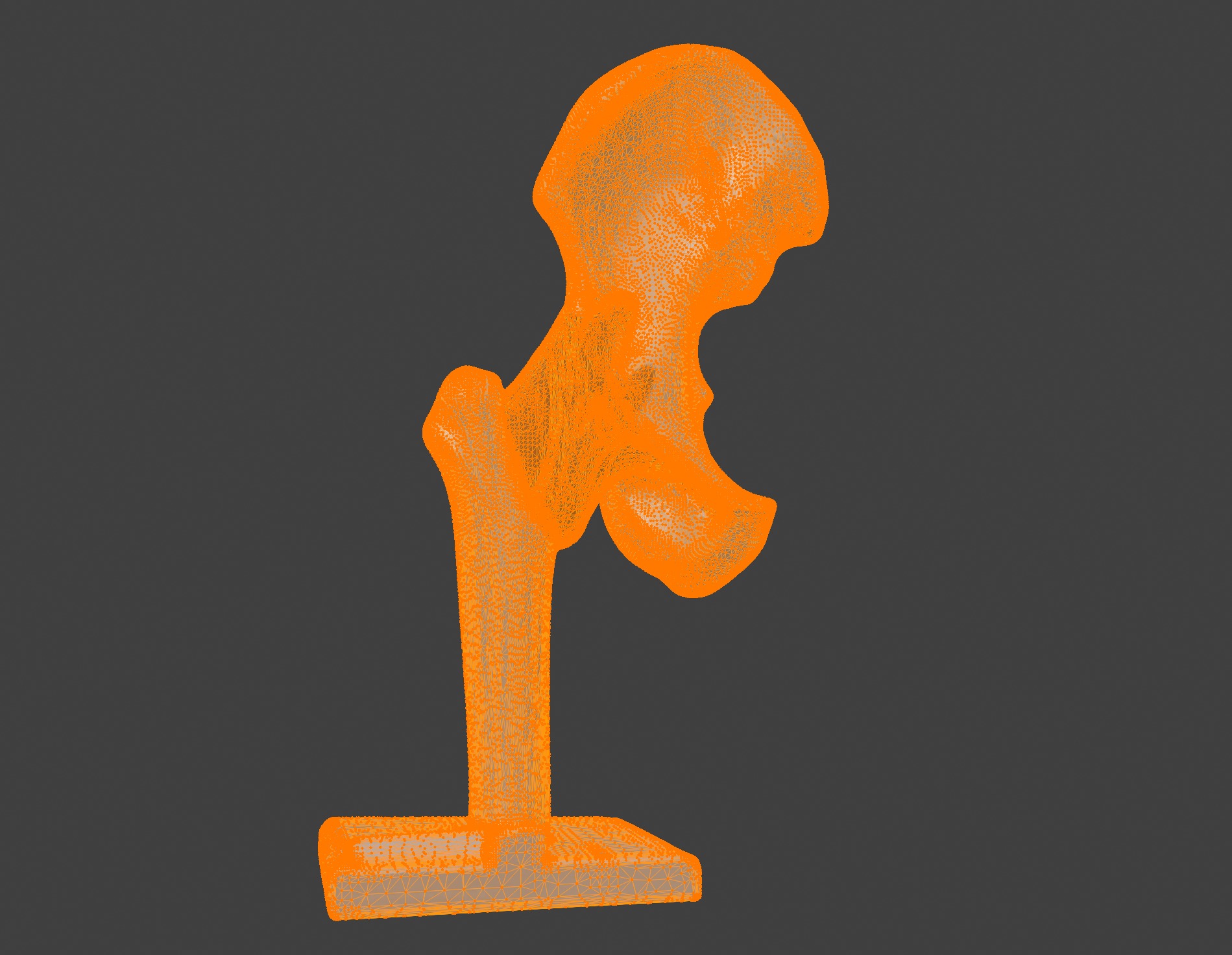
3D Model formats
Format limitations
- Collada (.dae)15.8 MB
- glTF (.gltf, .glb)4.36 MB
- OBJ (.obj, .mtl) (2 files)10.1 MB
- Blender (.blend)37.7 MB
- Autodesk FBX (.fbx)3.56 MB
3D Model details
- Ready for 3D Printing
- Publish date2025-01-15
- Model ID#5784508
Similar Models

Users who bought this item also bought...
