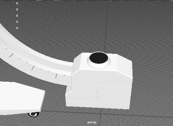
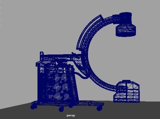
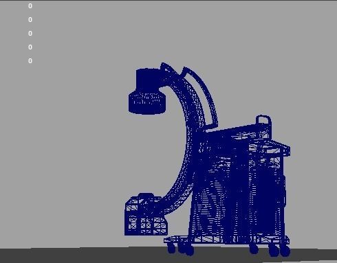

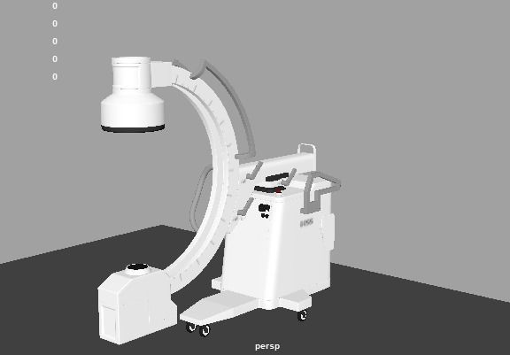
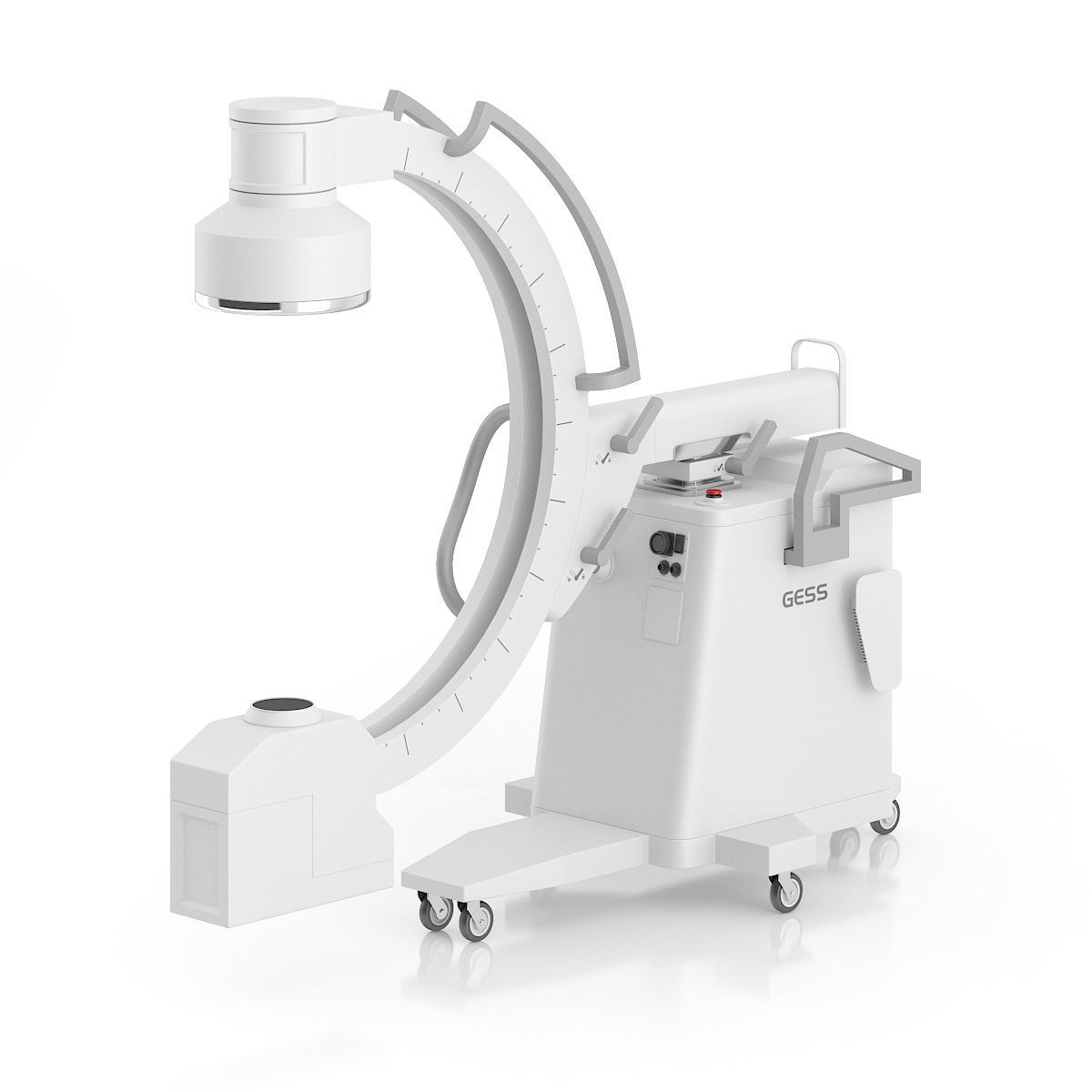
X ray Machine 3D model
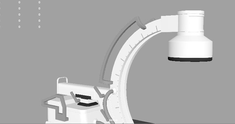
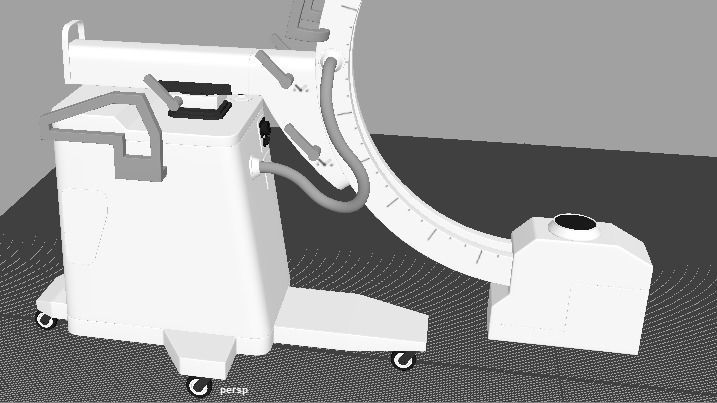
The X-ray Machine is a sophisticated medical imaging device that utilizes X-ray radiation to produce detailed images of the internal structures of the body. It is an essential tool in diagnostic medicine, allowing healthcare professionals to visualize bones, organs, and tissues to detect and diagnose various medical conditions.
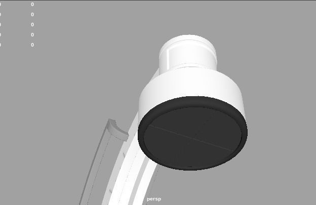
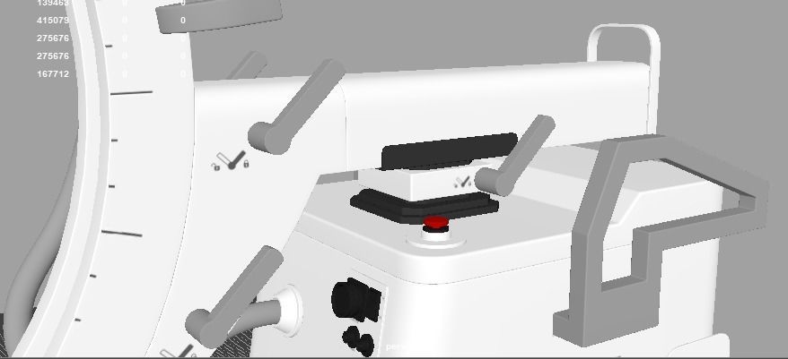
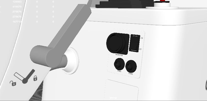
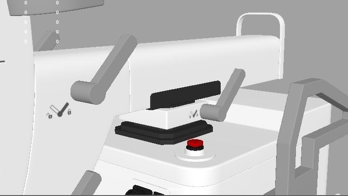
The X-ray machine consists of several components, including a control panel, X-ray tube, and a detector. The control panel allows the operator to adjust settings such as exposure time and radiation intensity. The X-ray tube emits a controlled dose of X-ray radiation, which passes through the body and interacts differently with various tissues. The detector captures the X-ray beams that pass through the body and converts them into digital images for interpretation.
During an X-ray procedure, the patient is positioned between the X-ray machine and the detector. The machine emits a focused beam of X-rays that penetrates the body and creates an image on the detector. The resulting X-ray image shows the internal structures, such as bones, organs, and soft tissues, allowing healthcare professionals to identify fractures, tumors, infections, and other abnormalities.
X-ray imaging is widely used in various medical specialties, including orthopedics, dentistry, cardiology, and pulmonary medicine. It is a valuable tool for diagnosing fractures, dislocations, lung diseases, dental problems, and evaluating the condition of organs such as the heart, lungs, and abdomen.
One of the significant advantages of X-ray imaging is its speed and non-invasiveness. X-rays can be obtained quickly, making it an efficient diagnostic tool in emergency situations. Additionally, X-ray imaging is generally safe, and the benefits of obtaining vital diagnostic information usually outweigh the minimal risks associated with the low levels of radiation exposure.
The interpretation of X-ray images is performed by radiologists, who are specialized physicians trained in diagnosing medical conditions through medical imaging. The radiologist analyzes the X-ray images and provides a detailed report to the referring healthcare provider, who will use the information to guide treatment decisions.
It is important to note that X-ray imaging has its limitations. It primarily visualizes dense structures, such as bones, but may have limited contrast in visualizing soft tissues. In certain cases, additional imaging techniques, such as computed tomography (CT) or magnetic resonance imaging (MRI), may be required for a more comprehensive evaluation.
The X-ray machine plays a crucial role in modern healthcare by providing valuable diagnostic information quickly and efficiently. Its use in medical facilities, clinics, and hospitals helps healthcare professionals make accurate diagnoses, plan treatments, and monitor patient progress.

3D Model formats
Format limitations
- Autodesk FBX (.fbx)41.1 MB
- Autodesk Maya (.ma, .mb)23.2 MB
- OBJ (.obj, .mtl) (2 files)48.4 MB
3D Model details
- Publish date2023-06-18
- Model ID#4579065
- Animated
- Rigged
- VR / AR / Low-poly
- PBR
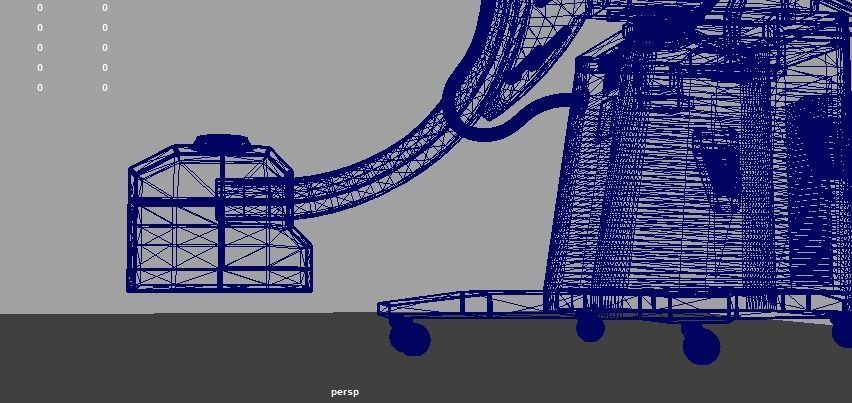
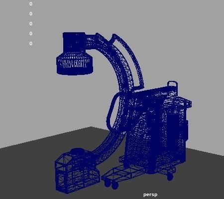
- Geometry Polygon mesh
- Polygons 275,676
- Vertices 139,463
- Textures
- Materials
- UV Mapping
- Unwrapped UVs Non-overlapping
- Plugins used
- Ready for 3D Printing
Similar Models